


清代红绿彩瓷器无损分析研究
[潘秋丽1  , 邵金发
, 邵金发1 , 李融武2 , 程琳1, * , 王荣1 ]
, 邵金发, 王荣|
|


作者简介: 潘秋丽, 1972年生, 北京师范大学核科学与技术学院实验师 e-mail: pql@bnu.edu.cn
红绿彩瓷器是我国陶瓷史上一种重要的釉上彩瓷器, 分析其彩料的化学组成和物相结构对其烧制工艺的研究有很重要的意义。 但由于瓷器表面的不平整和彩料分布的不均匀性, 导致其不满足传统的1 mm×10 mm线光源的X射线衍射仪对样品的测试要求。 而毛细管聚焦的X射线衍射仪采用点光源的方式照射样品, 毛细管X光透镜对Cu-Kα的能量有高达3个数量级的放大倍数, 同时具有低的发散度, 能实现样品直径100 μm的微区和直径3 mm的常规X射线衍射分析, 非常适合古陶瓷类样品矿物结构的无损分析的研究。 因此, 应用毛细管聚焦的微束X射线荧光谱仪和毛细管聚焦的X射线衍射谱仪对江西景德镇出土的清代红绿彩瓷的白釉和釉上彩料的化学成分和物相结构进行分析, 并对红绿彩瓷彩料中2 mm×2 mm感兴趣区域内多元素分布和矿物相的分布进行了二维扫描分析。 结果表明, Cu为绿彩的主要着色元素, 在绿彩中的含量为0.02%, 部分以Pb8Cu(Si2O7)3(PDF 31-0464)晶相形式存在; Fe为红彩的主要着色元素, Fe含量为1.63%, 部分Fe元素以Fe2O3(PDF 47-1409)的晶相形式存在; 其中Pb在绿彩和红彩中的含量分别为41.49%和6.29%, 其主要作用是使彩料的熔点降低, 部分Pb在700~800 ℃的烧制过程中与Cu元素和Si元素相结合以Pb8Cu(Si2O7)3(PDF 31-0464)晶相形式存在。 从扫描区域内的元素分布图和晶相分布图可以看出, 彩料原料中着色元素Cu和Fe的矿物晶相与Cu和Fe的元素分布不一致, 表明原料中原有的Cu和Fe的矿物晶相在烧制过程中基本上都消失了, 仅剩余或生成部分Fe2O3晶相; 白釉中存在莫来石晶相, 说明白釉是在高温下烧制而成; 其中Pb8Cu(Si2O7)3晶相的形成温度在750 ℃左右, 因此可以进一步说明清代红绿彩的绿彩料是在低温下烧制而成。 以上结果说明, 毛细管聚焦的微束X射线荧光和毛细管聚焦的微束X射线衍射谱仪在文物的科技研究中有着重要的应用前景。
The red and green colored porcelain is an important overglaze colored porcelain in the history of Chinese ceramics. It is significant to analyze the chemical composition and phase structure of the colored pigments to study the process of fired technologies. However, the traditional X-ray diffractometer with a 1 mm×10 mm line source is not easy to measure the porcelain sample of uneven surface and unevenly distributed color material. The capillary-focused X-ray diffractometer has a point X-ray beam to irradiate the sample. The capillary X-ray lens has a magnification of up to 3 orders of magnitude to the energy of Cu- Kα and has a low divergence of the X-ray beam. Therefore, it can measure the sample with a diameter of 100 micrometers and the conventional X-ray diffraction analysis of a sample with diameter of 3 mm, which is very suitable for the non-destructive analysis of the mineral structure of ancient ceramic samples. This article reports the results of the chemical composition and phase structure of the white glaze and red and green-colored pigments in the Qing Dynasty unearthed in Jingdezhen, Jiangxi province by the capillary focused microbeam X-ray fluorescence spectrometer, and the capillary focused X-ray diffraction spectrometer developed by our laboratory. It carried out the two-dimensional scanning analysis of the distribution of multi-element and mineral phases in the interest region (2 mm×2 mm) of the red and green colored porcelain. The results show that at first, Cu is the main coloring element of green colored pigments and the concentration of Cu in green colored pigments is 0.02%. Moreover, parts of Cu exist in the form of the Pb8Cu(Si2O7)3 crystal phase. Secondly, Fe is the main coloring element of red-colored pigments, and the contractions of Fe in red-colored pigments is 1.63%, and parts of Fe exist in the form of the Fe2O3 crystal phase; Thirdly, the concentrations of Pb in green and red-colored pigments is 41.49% and 6.29%, respectively. The main function of Pb is to lower the melting point of the colored pigments, and parts of the Pb combined with Cu and Si during the firing process at 700~800 ℃ to exist in the form of Pb8Cu(Si2O7)3 crystal phase. From the element 2D mapping and crystal phase mapping, it is obvious that the crystal phase of the coloring elements Cu and Fe in the colored pigments is inconsistent with the element distribution of Cu and Fe. It indicates that the Cu and Fe mineral crystal phases in the raw materials may disappear during the firing process, and the Fe2O3 crystal phases have been left or generated during the process of firing or cooling. The mullite crystal phase in the white glaze indicates that the white glaze fired at a high temperature. However, the formation temperature of the Pb8Cu(Si2O7)3 crystal phase is only about 750 ℃, which can further explain that the red and green colored pigments were fired at low temperatures. In general, the capillary-focused microbeam X-ray fluorescence spectrometer and the capillary-focused microbeam X-ray diffraction specrometer could have important application prospects in cultural relics’ scientific and technological research.
古陶瓷是中华文化重要的文化遗产, 蕴藏着中国古代先进的科学技术和艺术成就。 古陶瓷的化学成分和物相结构的分析对研究其原料、 配方和烧制工艺有很重要的意义[1, 2, 3]。 红绿彩瓷是一种釉上彩瓷, 是在白瓷的基础上用彩料装饰后再入窑在低温下烧制而制成[4]。 红绿彩瓷的研究历来受到古陶瓷研究者的关注[5]。 近年来, 随着红绿彩瓷器的大量发现, 古代红绿彩瓷的科学研究取得了一些进展[6]。 但由于红绿彩瓷彩料经过700~800 ℃的温度烧制, 彩料中部分矿物结构受到破坏。 同时, 由于瓷器表面不平整或具有较大弯曲度, 而常规X射线衍射仪采用1 mm× 10 mm线光源并且具有较高的发散度, 无法满足古陶瓷物相结构的无损分析的需求[6]。 毛细管X光透镜是一种X射线聚焦器件, 能将点光源激发出X射线束聚焦成直径几十~几百微米的束斑, 同时将X射线束的强度提高2~3个数量级, 能实现彩料微区的化学成分和元素分布的分析[7]。 此外, 毛细管X光透镜对Cu-Kα 具有最高的传输效率, 聚焦后的X射线束的强度提高3个数量级, 能实现样品微区的X射线衍射分析[8], 是一种理想的无损分析古陶瓷文物的化学成分和物相结构的分析方法。
采用本实验室台式微束X射线荧光谱仪对红绿彩瓷器彩料进行微区的点分析和二维元素分布分析[9]。 台式微束X射线荧光谱仪采用德国Rö ntgen公司的微焦斑X射线管(Mo靶, 焦斑: 30 μ m× 30 μ m)结合毛细管会聚X光透镜(在Mo-Kα 能量处焦斑直径为31 μ m)集成焦斑直径为31 μ m的高强度的微束X射线源; Amptek公司SDD(Silicon Drift Detector)X射线探测器(有效探测面积25 mm2, 5.9 keV能量处能量分辨率为145 eV)和PX5多道分析器; 高精度激光位移传感器实时矫正因样品不平整造成的位置偏移以及用于样品微区精确定位的CCD相机。
样品为江西省文物研究所提供的清代红绿彩瓷的残片(图1), 选取图1中A(白釉)、 B(红彩)、 C(绿彩)进行微区的元素组成分析。 实验设定, X射线管电压40 kV, 电流0.6 mA, 探测活时间300 s。 样品A(白釉)、 B(红彩)、 C(绿彩)三点的微束X射线荧光能谱如图2所示, 彩料中各元素化学成分采用基本参数法进行定量分析, 所得的数据如表1所示。
 | 图1 清代红绿彩瓷残片测量点与二维扫描区域 (2 mm× 2 mm)图Fig.1 The picture of a piece of red and green porcelain in Qing Dynasty and its scanned area |
 | 图2 白釉、 红彩和绿彩的微束能量色散X射线荧光谱Fig.2 μ -EDXRF spectra of white glaze and colored pigments |
| 表1 白釉、 红彩和绿彩的化学成分(质量分数, %) Table 1 Chemical compositions of white glaze and colored pigments (mass fraction, %) |
为了进一步探究清代红绿彩瓷彩料的元素与晶相的分布, 选取如图1所示的2 mm× 2 mm区域扫描, 对该区域进行μ -EDXRF和μ -XRD两种方式的二维扫描分析。 进行μ -EDXRF二维扫描分析时, X射线管电压为40 kV, 电流为0.6 mA, 微束X射线束斑为31 μ m(在Mo-Kα 能量处), 扫描步距为30 μ m, 每个点探测时间为1.5 s, 扫描数据经软件处理得到如图3所示的元素分布图。
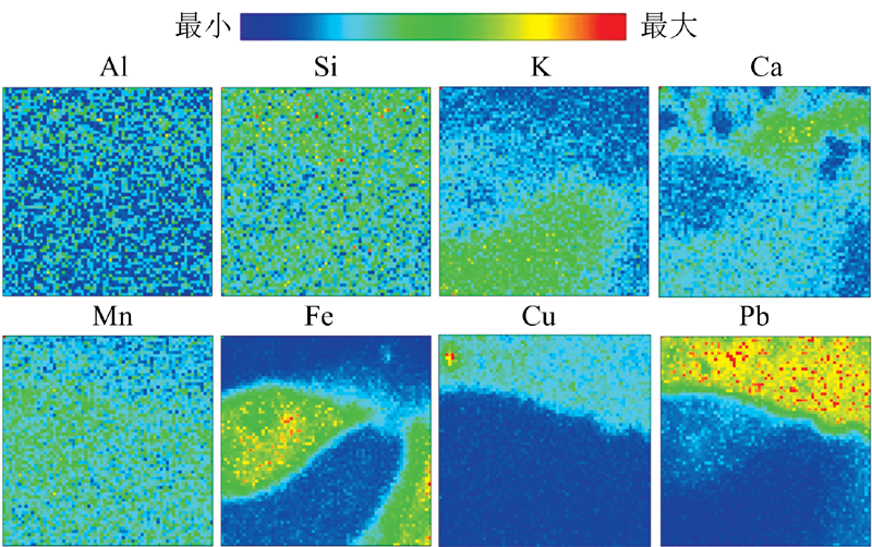 | 图3 扫描区域Pb, K, Fe, Ca, Cu, Al, Mn, Si元素的分布Fig.3 Elemental distributions of Pb, K, Fe, Ca, Cu, Al, Mn, Si in scanning areas |
X射线衍射分析采用本实验室毛细管聚焦的X射线衍射仪[10], 用Cu靶点光源X射线管(1 mm× 1 mm, 最大功率2 600 W)配备Ni吸收片和平行束X光透镜集成Cu-Kα 的平行束X射线源, 束斑直径约为3 mm; SDD X射线探测器(有效面积为25 mm2, 5.9 keV能量处能量分辨率为130 eV)和PX5单道脉冲分析器和间距为100 μ m的平行狭缝组组成X射线探测器系统; 采用θ -2θ 扫描模式, 角分辨优于0.01° 。 利用X射线衍射仪对清代红绿彩瓷残片(图1)中A(白釉), B(红彩), C(绿彩)进行XRD分析[10]。 测量时, Cu靶X射线管电压为40 kV, 电流为40 mA, 角扫描步距为0.15° , 2θ 探测范围为10° ~90° , 每步探测时间为5 s, XRD谱图如图4。
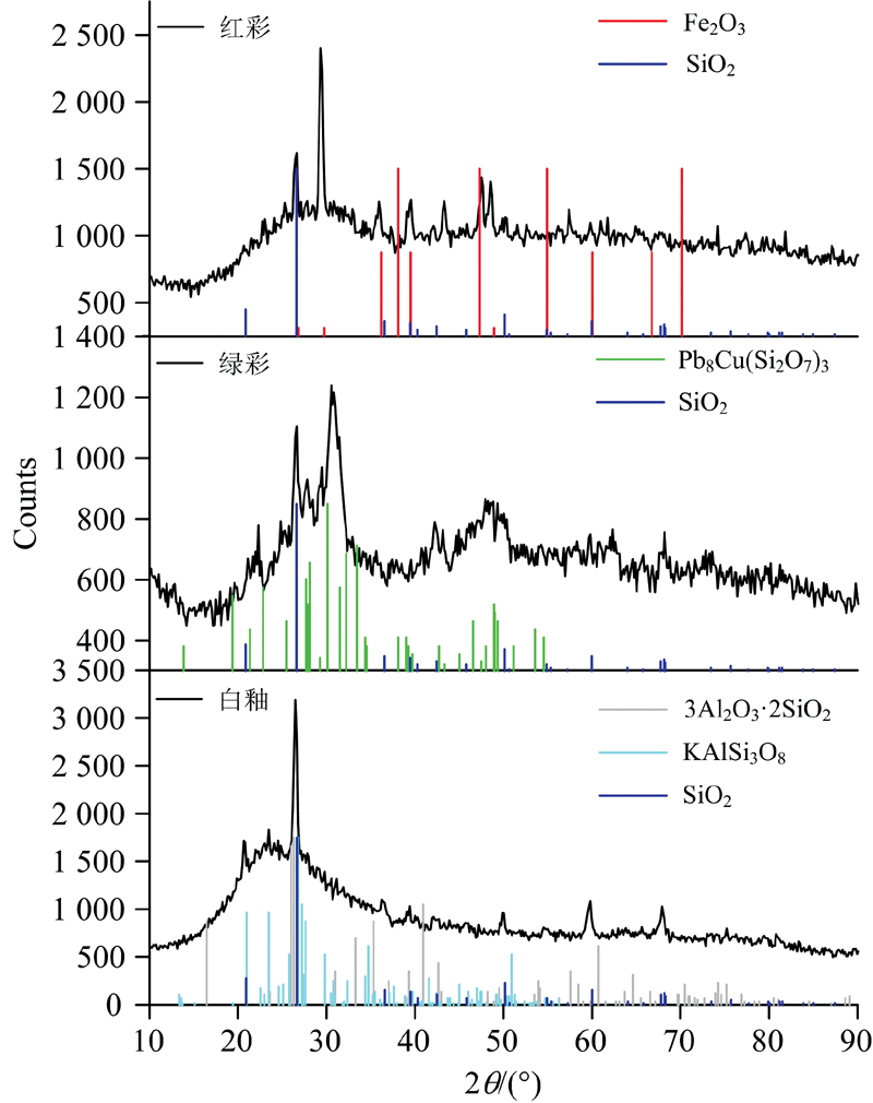 | 图4 白釉、 红彩和绿彩的X射线衍射谱图Fig.4 XRD patterns of white glaze and colored pigments |
从图4中可以看出, A点白釉XRD谱图在15° ~35° 之间出现一个驼峰, 这是白釉在高温烧制过程中形成的非晶相所致; 同时, 经过对比ICCD PDF卡, A点白釉中主要存在的晶相为钾长石KAlSi3O8(PDF 25-0618)、 石英SiO2(PDF 46-1045)和莫来石3Al2O3· 2SiO2(PDF 15-0776)等; B点红彩中主要存在的晶相为Fe2O3(PDF 47-1409)和石英SiO2(PDF 46-1045)等; C点绿彩中主要存在的晶相为Pb8Cu(Si2O7)3(PDF 31-0464)等。
为验证XRD实验结果的准确性, 采用法国Horiba Jobin Yvon公司的LabRAM Aramis型号激光共聚焦微拉曼散射光谱仪, 选用波长为532 nm(YAG固体激光器)的激光作激发源, 对红绿彩瓷的A点白釉、 B点红彩、 C点绿彩进行了测试, 得到如图5所示的拉曼光谱图。 由图4可知, A点白釉和C点绿彩未出现明显的峰位; B点红彩中出现了Fe2O3晶相信号, 与XRD测试结果基本一致。 通过激光拉曼光谱与毛细管聚焦的X射线衍射仪测量结果的对比可知, 本实验室研发的毛细管聚焦的X射线衍射仪比拉曼光谱仪更适合于古陶瓷类样品的无损分析。
 | 图5 白釉、 红彩和绿彩的拉曼光谱图Fig.5 Raman spectra of white glaze and colored pigments |
为明确红绿彩瓷中矿物晶相在彩料中的分布, 在图1中2 mm× 2 mm的区域里, 用微束X射线衍射仪进行μ -XRD二维扫描分析[11]。 微束X射线束斑为100 μ m(在Cu-Kα 能量处), 扫描步距为100 μ m, 该区域被划分为21× 21个被测试点。 每个点的测量条件为: X射线管电压为30 kV, 电流为0.5 mA, 2θ 探测范围为24.5° ~30.5° , 步距角为0.3° , 每步探测活时间为0.8 s。 由此得到的扫描总谱经数据处理得到的晶相分布图如图6所示。
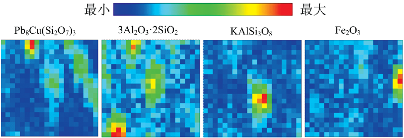 | 图6 扫描区域Pb8Cu(Si2O7)3, 3Al2O3· 2SiO2, KAlSi3O8, Fe2O3的晶相分布Fig.6 Phase mappings of Pb8Cu(Si2O7)3, 3Al2O3· 2SiO2, KAlSi3O8, Fe2O3 in scanning areas |
在红绿彩瓷的烧制过程中, 彩料的原料发生熔融, 并发生一系列物理化学变化。 在随后的冷却过程中, 随着温度的降低, 发生分相和结晶, 导致瓷器釉和彩料中元素分布不均匀和某些矿物相的析出。 从红绿彩瓷器已有的分析结果结合以上的实验结果可以得知, Cu为绿彩的主要着色元素, 在绿彩中的含量为0.02%, 部分以Pb8Cu(Si2O7)3(PDF 31-0464)晶相形式存在; Fe为红彩的主要着色元素, Fe含量为1.63%, 部分Fe元素以Fe2O3(PDF 47-1409)的晶相形式存在; 其中Pb在绿彩和红彩中的含量分别为41.49%和6.29%, 其主要作用是使彩料的熔点降低, 部分Pb在700~800 ℃的烧制过程中与Cu元素和Si元素相结合以Pb8Cu(Si2O7)3(PDF 31-0464)晶相形式存在。 从扫描区域内的元素分布图和晶相分布图可以看出, 彩料原料中着色元素Cu和Fe的矿物晶相与Cu和Fe的元素分布不一致, 表明原料中原有的Cu和Fe的矿物晶相在烧制过程中基本上都消失了, 仅剩余或生成部分Fe2O3晶相; 白釉中存在莫来石晶相, 说明白釉是在高温下烧制而成; 其中Pb8Cu(Si2O7)3晶相的形成温度在750 ℃左右, 因此可以进一步说明清代红绿彩的绿彩料是在低温下烧制而成。
对清代红绿彩瓷残片进行了微束X射线荧光分析和X射线衍射分析, 结果表明白釉中存在钾长石(KAlSi3O8)和莫来石(3Al2O3· 2SiO2)晶相; 彩料中存在Fe2O3 和Pb8Cu(Si2O7)3晶相, 其中Pb8Cu(Si2O7)3晶相的存在表明其彩料是在750 ℃左右烧制而成的。 此外, 也说明毛细管聚焦的X射线荧光谱仪和毛细管聚焦的X射线衍射谱仪在文物的科技研究中有重要的应用前景。
| [1] |
|
| [2] |
|
| [3] |
|
| [4] |
|
| [5] |
|
| [6] |
|
| [7] |
|
| [8] |
|
| [9] |
|
| [10] |
|
| [11] |
|