青碧与青玉的红外光谱特征及意义
[任建红1  , 施光海
, 施光海1, * , 张锦洪2 , 袁野1 , 高孔1 , 王美丽1 , 李新岭3 , 龙楚4 ]
, 施光海, 张锦洪|
|


作者简介: 任建红, 女, 1990年生, 中国地质大学(北京)博士研究生 e-mail: renjhwang@163.com
青碧为碧玉系列中外观类似青玉的称呼。 二者虽外观相似, 但青碧价格高很多, 故市场上出现了以青玉充青碧售卖的现象。 此外, 一些出土玉文物中也出现了这类外观的玉石材质, 但无法准确判别其类型。 这使得快速准确鉴别二者有十分重要的意义。 采用紫外-可见光谱、 傅里叶变换红外光谱和电子探针分析方法, 给出了青碧和青玉样品的谱学特征和矿物的化学组成等特征, 并进行了对比分析。 二者在紫外-可见反射光谱上没有明显差异, 然而, 二者的红外光谱特征存在可识别的差异。 为了探讨出更有效的鉴别特征, 采用了反射和透射两种方法来获取红外光谱。 二者的红外光谱总体上一致, 有以下可区分差异特征: 青碧的红外反射光谱中出现了青玉光谱中未出现的1 050和1 018 cm-1附近吸收峰、 肩峰及411 cm-1附近宽肩峰; 青玉的红外透射光谱中出现了青碧光谱中未出现的453 cm-1附近肩峰和401 cm-1附近吸收峰。 以上可作为快速鉴别青碧和青玉的谱学特征标志。 红外透射光谱经朗伯-比尔定律( A=log(1/ T))转换后, 在3 674, 3 661和3 643 cm-1附近处的OH伸缩振动谱带的强度与M1, M3位的Mg及Fe2+含量有很好的相关性。 利用以上二者关系计算的Mg(M1+M3)#(在M1和M3位的Mg/(Mg+Fe2+))值可用于鉴别青碧和青玉。 青碧的Mg(M1+M3)#值为0.871~0.892, 小于青玉0.927~0.949。 另外, 电子探针分析结果显示青碧和青玉的化学成分存在一定差异: 青碧Mg含量(a.p.f.u)为4.45~4.53, 比青玉的4.66~4.78小; 青碧Fe2+含量为0.28~0.49, 大于青玉的0.10~0.23。 但部分青碧和青玉间的Mg和Fe2+含量差异不大, 说明红外光谱差异除了与成分有一定的关联性外, 可能还与结晶时的物理化学条件有关(青碧和青玉的成因类型分别为超基性岩型和白云质大理岩型)。 以上红外光谱识别特征不仅在鉴别青碧和青玉上具有重要的宝石学意义, 还在古代玉制品源区的判别、 产状分析等方面具有潜在的应用价值。
, SHI Guang-hai, ZHANG Jin-hongGrayish green nephrite is named for a kind of nephrite belonging to green nephrite type, but with appearance similar to gray nephrite. Although their appearance is similar, the price of grayish green nephrite is much higher than that of gray nephrite. Thus a phenomenon appears that some dealers tell their consumers green nephrite while selling gray nephrite. In addition, some jade materials with such appearance appear in some unearthed jade artifacts, but their types can not be accurately identified. This makes it particularly important to quickly and accurately identify grayish green nephrite and gray nephrite. In this study, representative grayish green nephrite and gray nephrite samples were investigated using ultraviolet-visible spectroscopy, Fourier transform infrared spectroscopy and electron microprobe analysis, and all the characteristics were yielded. By comparing the features between them, it can be found that there is no significant difference in the UV-Vis reflection spectra of both types of samples. However, the differences in the infrared spectra of them are recognizable. In order to explore more effective identification features, the reflection and transmission methods were used to obtain infrared spectra. The infrared spectra of both types of samples were generally the same, with the following distinguishable differences. The peak or shoulder around 1 050 and 1 018 cm-1 and the broad shoulder near 411 cm-1 occur in the reflection spectra of grayish green nephrite which do not appear in those of gray nephrite. The shoulder around 453 cm-1 and the peak near 401 cm-1 exhibit in the transmission spectra of the gray which do not exist in those of the grayish green. The above findings can be used as spectral characteristics to identify grayish green nephrite and gray nephrite. The intensity of the OH stretching vibration bands at 3 674, 3 661 and 3 643 cm-1 after Beer-Lambert Law transformation of the infrared transmission spectra and the Mg and Fe2+ content in the M1 and M3 sites are well correlated. The Mg(M1+M3)# (Mg/(Mg+Fe2+) in the M1 and M3 sites) ratio calculated by the relationship between the above two can be used to distinguish between grayish green nephrite and gray nephrite using their infrared transmission spectra. Mg(M1+M3)# ratio in grayish green nephrite (0.8710.892) is smaller than that of gray nephrite (0.9270.949). Moreover, the result of electron microprobe analysis showed that there are some differences in chemical composition between them. Mg content in grayish green nephrite (4.454.53) is less than that of gray nephrite (4.664.78), and Fe2+ content in the grayish green (0.280.49) is larger than that of the gray (0.100.23). However, Mg and Fe2+ content between them are not much different from each other, suggesting that the difference in infrared spectra may be related to the physicochemical conditions during crystallization besides having a certain correlation with the composition (the genetic types of grayish green nephrite and gray nephrite are ultrabasic rock type and dolomitic marble type, respectively). The above infrared spectrum identification features not only have important gemological significance for identification of grayish green nephrite and green nephrite, but also have potential application value for discriminating origin and analyzing occurrence of some ancient jades with the similar appearance to the studied nephrites.
软玉是以透闪石-阳起石为主要组成的隐晶致密矿物集合体[1, 2, 3], 按其成因, 主要划分为两类: 一类与超基性岩有关, 另一类与白云质大理岩有关。 在商业贸易中, 软玉常以颜色进行分类, 如白玉、 青白玉、 青玉、 碧玉、 墨玉等, 其中碧玉的成因类型几乎都与超基性岩有关。 在碧玉系列中, 有时会出现少量颜色相对较浅的碧玉, 其外观和与白云质大理岩相关的青白玉-青玉系列中的一些青玉十分相似。 本工作将碧玉中青色者定名为青碧, 用来与青玉区分。 尽管二者外观相似, 但青碧由于稀少, 有“ 鸭蛋青、 雪山碧玉” 等商业名称, 价格自然比青玉高得多, 从而在市场上出现了将青玉充当青碧进行售卖的现象。 因此, 青碧和青玉的鉴别成为市场对软玉研究提出的一项新任务。
在实际鉴定工作中, 二者的区分较为困难。 对于一般消费者而言, 很难判断商家标注青碧或青玉品种的可靠性, 就是经验丰富的专业人士, 单从颜色、 结构等外观特征, 也没有十分的把握来辨别青碧和青玉。 此外, 考古出土的文物中也出现了这类外观相似但不辨类型的玉材。 这使得通过简单可靠的分析方法找到青碧和青玉的鉴别标志显得尤为重要。 近年来, 谱学方法已被广泛应用于宝玉石研究, 如物相鉴定、 阳离子含量检测和地质成因探究等[3, 4, 5, 6, 7]。 然而, 目前为止, 针对青碧和青玉的谱学研究相对较少, 还未见到公开报道。
以外观相似的青碧和青玉样品为研究对象, 采用紫外-可见光谱、 傅里叶变换红外光谱和电子探针分析方法, 对二者的谱学特征和矿物的化学组成等特征进行了系统性对比研究, 找出了青碧和青玉的谱学鉴别标志, 并探讨了红外光谱可能的鉴定意义。
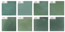
从外观特征相似的大量青碧和青玉中选取研究样品(图1)。 其中, 青碧样品采于青海祁连县托来南山和俄罗斯东萨彦岭7号矿点, 为超基性岩型软玉。 青玉样品采于新疆且末县镍旺和于田县戚家坑矿点, 为白云质大理岩型软玉。 每个矿点选取两块代表性样品。 研究样品均呈浅青色, 结构致密细腻, 其描述见表1。
 | 图1 青碧和青玉样品的切片照片 RU-1, RU-2, QH-1和QH-2为青碧样品; YT-1, YT-2, QM-1和QM-2为青玉样品; 样品长宽均为1 cmFig.1 Slice photograph of grayish green nephrite and gray nephrite RU-1, RU-2, QH-1 and QH-2 are grayish green nephrite; YT-1, YT-2, QM-1 and QM-2 are gray nephrite; the length and width of samples are 1 cm |
| 表1 青碧和青玉样品描述 Table 1 Descriptions of grayish green nephrite and gray nephrite |
使用中国地质大学(北京)珠宝学院的UV-3600型紫外-可见分光光度计, 获得了样品的紫外-可见反射光谱。 测试条件: 波长范围200~800 nm, 采样间隔0.5 nm。 还使用了珠宝学院的BRUKER TENSOR 27型傅里叶变换红外光谱仪, 分别采用反射法和透射法获得了样品的红外光谱。 反射法测试条件: 扫描范围2 000~400 cm-1, 扫描32次, 分辨率4 cm-1。 透射法测试条件: KBr压片, 扫描范围4 000~400 cm-1, 扫描16次, 分辨率4 cm-1。 样品矿物成分测试在中国地质大学(北京)电子探针实验室的EMPA-1720型电子探针仪上完成。
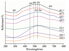
青碧和青玉的紫外-可见反射光谱中均出现280, 450, 490, 530和660 nm附近吸收峰或肩峰, 仅俄罗斯青碧(RU-1和RU-2)出现了其他样品光谱中未出现的510 nm弱峰和550 nm肩峰(图2)。 可见, 二者的紫外-可见反射光谱特征基本一致, 故目前无法根据紫外-可见反射光谱特征对青碧和青玉进行有效的区分。
 | 图2 青碧和青玉样品的紫外-可见反射光谱Fig.2 UV-Vis reflection spectra of grayish green nephrite and gray nephrite |
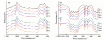
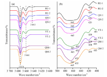
红外反射和透射光谱测试结果表明: 青碧和青玉的红外光谱特征基本一致, 但在一些峰位、 峰强或峰形上存在可识别的差异(图3和表2)。
 | 图3 青碧和青玉样品的红外反射光谱(a)和透射光谱(b)Fig.3 Infrared reflection spectra (a) and transmission spectra (b) of grayish green nephrite and gray nephrite |
| 表2 青碧和青玉样品红外吸收峰的位置及归属 Table 2 Location and attribution of infrared absorption peaks of grayish green nephrite and gray nephrite |
2.2.1 红外反射光谱差异
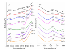
(1)在1 100~1 000 cm-1范围内, 青碧出现四个明显的吸收峰、 肩峰, 分别位于1 092, 1 050, 1 040和1 018 cm-1附近, 部分样品在1 047 cm-1处为肩峰(RU-1和RU-2), 部分样品在1 018 cm-1处为肩峰(QH-1和QH-2)。 青玉在该范围只出现两个强吸收峰, 分别位于1092和1040 cm-1附近[图4(a)]。
 | 图4 青碧和青玉样品在1 1001 000 cm-1(a)和 440400 cm-1(b)范围内的红外反射光谱Fig.4 Infrared reflection spectra of grayish green nephrite and gray nephrite in the range of 1 1001 000 cm-1 (a) and 440400 cm-1 (b) |
(2)在440~400 cm-1范围内, 青碧含有417 cm-1附近吸收峰和411 cm-1附近宽肩峰, 而青玉仅有417 cm-1附近吸收峰。 比较来看青碧两吸收峰峰形较青玉宽缓[图4(b)]。
2.2.2 红外透射光谱差异
(1)在3 700~3 600 cm-1范围内, 青碧存在三个吸收峰, 分别为3 674 cm-1强吸收峰、 3 661 cm-1中等吸收峰和3 643 cm-1附近弱吸收峰, 而青玉仅存在3 674 cm-1强吸收峰和3 661 cm-1中等吸收峰[图5(a)]。
 | 图5 青碧和青玉样品在3 7003 600 cm-1(a)和500400 cm-1(b)范围内的红外透射光谱(图5(a)中光谱经基线校正和高斯拟合)Fig.5 Infrared transmission spectra of grayish green nephrite and gray nephrite in the range of 3 7003 600 cm-1 (a) and 500400 cm-1 (b) (spectra in the fig.5(a) are calibrated by baseline and Gaussian) |
(2)在500~400 cm-1范围内, 青碧具有465和420 cm-1两个吸收峰, 而青玉含有三个吸收峰和一个肩峰, 分别位于465, 453(肩峰), 420和401 cm-1附近[图5(b)]。
2.2.3 Mg(M1+M3)#值
透闪石-阳起石系列矿物的晶体化学式为Ca2(M12, M22, M3)5[Si4O11]2(OH)2, 其中M1~M3位通常被Mg和Fe2+占据, OH位于硅氧四面体组成的六方环的中心, 与两个M1位及一个M3位上的Mg和Fe2+相连。
前人认为不同类型软玉在M1和M3位的Mg/(Mg+Fe2+)值是不一样的[8]。 红外透射光谱中, 位于3 674, 3 661和3 643 cm-1附近处的OH伸缩振动谱带分别对应OH(MgMgMg)带、 OH(MgMgFe2+)带、 OH(MgFe2+Fe2+)带[图5(a)]。 红外透射光谱经朗伯-比尔定律(A=log(1/T), A表示吸光度, T表示透射率)转换后, 根据各带的强度、 占位阳离子种类及比率, 计算出M1和M3位的Mg和Fe2+含量及Mg/(Mg+Fe2+)#值, 分别记为Mg(M1+M3), Fe2+(M1+M3)及Mg(M1+M3)#(表3)。 数据显示, 青碧和青玉的Mg(M1+M3)#值分别介于0.871~0.892和0.927~0.949之间; 青碧明显小于青玉。
| 表3 青碧和青玉样品的Mg(M1+M3)#值 Table 3 Mg(M1+M3)# ratios in grayish green nephrite and gray nephrite |
青碧和青玉中矿物的主要成分为SiO2, MgO和CaO(表4), 其含量分别为57.79%~58.71%, 21.47%~23.58%, 12.60%~13.78%, 这些值与透闪石化学成分理论值十分接近。 该矿物的晶体化学式(基于23个氧原子计算)与透闪石理论分子式吻合, Mg#=Mg/(Mg+Fe2+)值均大于0.9。 以上分析结果表明, 二者的主要矿物组成均为透闪石。 另外, 二者Mg和Fe2+含量差异较大: 青碧Mg含量(a.p.f.u)为4.45~4.53, 小于青玉的4.66~4.78; 青碧Fe2+含量为0.28~0.49, 大于青玉的0.10~0.23。 然而, 部分青碧和青玉间的Mg和Fe2+含量差异并不明显(QH-2和YT-1)。
| 表4 青碧和青玉样品中矿物的电子探针分析结果 Table 4 Electron microprobe analyses for grayish green nephrite and gray nephrite |
青碧和青玉的红外光谱特征存在可分辨的差异, 这可能与其Mg和Fe2+含量及结晶时的物理化学条件不同有关。
透闪石晶体结构中的OH可与两个M1位和一个M3位的Mg和Fe2+成键, 出现四种组合方式, 分别为OH(MgMgMg), OH(MgMgFe2+), OH(MgFe2+Fe2+)和OH(Fe2+Fe2+Fe2+), 分别对应红外光谱中的3 674, 3 661, 3 645和3 625 cm-1附近谱带, 由于Fe2+的电负性比Mg强, 故使M— O键增强, 而O— H键减弱, 因而随着Fe2+含量的增加, 其逐渐置换Mg, OH伸缩振动谱带逐渐出现。 可能由于上述置换导致Si— O和M— O键的性质发生变化, 从而产生不同频率的振动。
电子探针分析结果显示, 青碧和青玉的Mg和Fe2+含量确实存在差异, 但部分样品间的Mg和Fe2+含量差异并不十分明显, 说明红外光谱差异不只与成分有关。 已知青碧产于超基性岩和基性火山岩的接触蚀变带, 而青玉产于中酸性侵入岩与白云质大理岩的接触带, 即二者形成时的温度、 压力、 pH值等物理化学条件差异较大, 故红外光谱差异可能还与结晶时的物理化学条件有关。
可见, 青碧和青玉在成分和结晶时的物理化学条件等方面存在的差异可能是二者红外光谱特征出现可分辨差异的主要原因。 故红外光谱对青碧和青玉的鉴别具有重要意义。 在考古学中也出现过类似外观的玉材, 红外光谱对确定其产状也具有非常重要的指示意义。
(1)青碧和青玉的紫外-可见反射光谱特征基本一致, 故目前无法根据紫外-可见光谱特征将青碧和青玉进行分别。
(2)青碧和青玉的红外反射光谱及透射光谱均存在可分辨的差异。 根据反射光谱中有无1 050和1 018 cm-1附近吸收峰、 肩峰及411 cm-1附近宽肩峰与透射光谱中是否存在453 cm-1附近肩峰及401 cm-1附近吸收峰可以快速鉴别青碧和青玉。
(3)根据红外OH伸缩振动谱带的强度、 占位阳离子种类及比率计算出青碧和青玉的Mg(M1+M3)#值分别介于0.871~0.892和0.927~0.949之间; 青碧明显小于青玉。 该值可作为鉴别标志。
(4)电子探针分析结果显示二者Mg和Fe2+含量存在差异: 青碧Mg含量小于青玉, 其Fe2+含量大于青玉。 但部分青碧和青玉间的Mg和Fe2+含量差异不大, 说明红外光谱差异与成分存在一定的关联性。 另外, 所研究的青碧和青玉结晶时的物理化学条件不同, 也可能导致二者的红外光谱存在差异。
(5)利用红外光谱差异特征可快速准确鉴别青碧和青玉, 这对古玉器地质源区的判别具有潜在的应用价值。
致谢: 感谢新疆维吾尔自治区玛纳斯县高彦海先生及刘强先生等在矿区考察及采样工作中给予的大力支持!
The authors have declared that no competing interests exist.
| [1] |
|
| [2] |
|
| [3] |
|
| [4] |
|
| [5] |
|
| [6] |
|
| [7] |
|
| [8] |
|