


窄带紫外LED照射对大鼠骨代谢的影响
[李云奇1  , 张宁
, 张宁2 , 陈德福3 , 邱海霞1 , 曾晶1 , 王娜5, * , 顾瑛1, 3, 4, * ]
, 张宁, 顾瑛]
|
|


背景:紫外线是维持骨骼健康的关键条件之一, 但由于传统的紫外线光谱较宽,在骨质疏松症领域并未引起足够的重视。 材料与方法: 在研究中, 建立了一种新设计的窄带光谱LED器件, 以研究紫外LED对骨质疏松大鼠骨代谢、 骨形态和皮肤的影响。 共设置了去卵巢大鼠(n=24)和去卵巢大鼠(n=36)。 然后将去卵巢大鼠分为假手术组(Sham,n=12)和检测模型组(Sham,n=12)。 将去卵巢大鼠分为LED照射组(LED,n=12)、 未处理组(OVX,n=12)或检测模型组(OVX,n=12)。 LED 辐照参数(0.8 mW·cm-2, 1 000 s,每周两次)。结果:与假手术组相比, 维生素D、 mRNA、 血清ALP、 血清25(OH)D3、 血清P1NP的表达明显升高, 血清PTH和血清TRAP的表达明显降低。 大鼠皮肤照射前后p53基因未见明显变化。结论:研究结果表明, 新型紫外 LED 器件照射可显着提高血液中 25(OH)D3 水平, 促进骨形成, 抑制骨吸收, 且对大鼠皮肤无不良影响。
Biography: LI Yun-qi, (1987—), attending doctor, The First Medical Center, Chinese PLA General Hospital e-mail: yunqi206@126.com
Background:Ultraviolet radiation is one of the key conditions to maintaining bone health, but it has not been paid enough attention to in the field of osteoporosis because of the wide traditional ultraviolet spectrum. With the development of LED technology, all kinds of narrowband LED spectra can be adjusted arbitrarily. Materials and methods: In this study, we established a newly designed LED device with a narrow band spectrum to investigate the effects of ultraviolet LED on bone metabolism, bone morphology and skin of osteoporotic rats. We have set up a total of unovariectomized rats (n=24) and ovariectomized rats (n=36). The unovariectomized rats were then divided into the sham operation group (Sham,n=12) and the detection model group (Sham,n=12). The ovariectomized rats were divided into either the LED irradiation group (LED,n=12), the no treatment group (OVX,n=12), or the detection model group (OVX,n=12). The LED Irradiation parameters (0.8 mW·cm-2, 1 000 s, twice a week).Results:Compared with the sham group, the expression of vitamin D mRNA, serum bone ALP, serum 25(OH)D3, and serum P1NP increased significantly, the serum PTH and serum TRAP decreased significantly. Conclusion:Our results show that irradiation with the new ultraviolet LED device can significantly increase the level of 25(OH)D3 in blood, promote bone formation and inhibit bone resorption without adverse effects on rat skin.
Osteoporosis is a systemic bone disease. With the continual increase in people's life expectancy, osteoporosis is also increasing year by year[1, 2, 3]. Among the complications associated with osteoporosis, fractures are the most serious. According to statistics, 10 million cases of osteoporotic fracture occur worldwide, seriously threatening people's health and quality of life. The combination of the prevalence of osteoporosis and the increasing cost of health care has created a significant public health issue[4, 5, 6]. The current osteoporosis treatment method is mainly drug therapy, such as the synthetic metabolic drug bisphosphate, which inhibits bone resorption of estrogen[7, 8, 9, 10]. Drugs such as these could improve bone microarchitecture and decrease fracture risk. However, these drugs have many limitations and toxic side effects that can affect how long patients can take them without risk[11, 12].
Ultraviolet radiation is a key condition that helps maintain bone health; approximately 80% of human body's vitamin D is synthesized from exposure to sunlight[13, 14, 15]. However, the amount of exposure to sunlight someone receives is greatly affected by altitude, latitude, season, weather conditions and the time of day, so without guidance, it is not easy to get sufficient vitamin D from exposure to sunlight. Previous studies have shown that fluorescent tubes containing UV radiation can help increase vitamin D synthesis in the skin, subsequently enhancing intestinal calcium absorption and thereby maintaining bone mass and reducing bone loss, thus reducing the incidence of fractures. However, due to the varying range of wavelengths emitted by fluorescent tubes, patients could be exposed to unnecessary and harmful wavelengths while simultaneously receiving therapeutic wavelengths, thus limiting the broad application of this approach for osteoporosis treatment[13, 14]. LED (light-emitting diode) lights are a new alternative light source. Currently, the applications of UV LED technology are mainly limited to the fields of disinfection, sterilization, and deodorization. Therefore, little is known about the possible value of this technology for treating osteoporosis.
Rapid improvement in semiconductor technology promotes the application expansion of nitride-based light-emitting diode (LED). The device operates in visible spectra and has been widely used in solid-state lighting (SSL) due to a list of advantages over traditional light sources, such as compact size, energy efficiency, low environmental impact, low cost, long lifetime and robustness. The benefits resulting from the transition from the conventional light sources to SSL have been witnessed, and the Nobel Prize in Physics 2014 was awarded to the scientists for the invention of efficient blue LED. Recent research progress has shown that the capability and potential application scenario of III-nitride-based LED are beyond basic illuminance and display. Since the spectrally pure emission at the desired wavelength of LED can be engineered by adjusting the element composition of the semiconductor material, the device attracts great attention and is a good candidate in wireless communication, bio-detection, curing, purification and disinfection etc[16, 17, 18, 19, 20]. AlGaN-based ultraviolet (UV) LED has already exhibited inactivation efficiency in fighting against severe acute respiratory syndrome coronavirus 2 (SARS-CoV-2). However, the effectiveness of UV irradiation in light therapy[21], especially in treating osteoporosis, needs to be studied.
Here, we report for the first time the comprehensive study of the effects of UV LED irradiation on bone metabolism in a rat osteoporosis model. This study aimed to provide an experimental basis for possible clinical application of UV LED devices in clinical treatment of osteoporosis.
The peak wavelength of the UV LED is 293 nm. Because of studies have shown that in all spectra, 290~300 nm UV wavelengths are most effective for vitamin D production in the equivalent model of human skin or human skin[25-26. The LED irradiation array was constructed according to the experimental desired irradiation area (30 cm2) for the experimental rats. The aluminum substrate was used to dissipate heat, a rapid pressing tool and a defined position block were installed for fixing, the upper seal and the 120 mm× 25 mm gap pressure plate were installed, and the exhaust fans were installed at both ends to keep the temperature constant [Figure 1(a)]. The reflectance of the box was 1.3, and the power density of the UV radiation was 0.8 mW· cm-2, delivered for a duration of 1 000 s, twice a week, for a total of 12 weeks. The energy density was selected according to the rats' minimum sub erythema dose for rats. The irradiation parameters was are listed in Table 1. The LED irradiation group received the UV LED irradiation treatment, while the sham operation group did not. After 12 weeks of irradiation, the rats in all groups were euthanized, and samples (right femur, liver and skin) were harvested.
| Table 1 Irradiation parameters |
60 healthy 3-month-old female SD rats (180~200 g) were purchased from the Experimental Animal Center of the General Hospital of the Chinese People's Liberation Army. The room temperature of the animal feeding environment was constant [(20± 2) ℃], the relative humidity was 40%, and the ventilation was adequate. Rats were housed with water ad libitum, in a 12:12 hour light-dark cycle with protection from UVB exposure until the end of the study. According to the principle of random grouping, the 60 osteoporotic rats were randomly divided into unovariectomized (n=24) and ovariectomized (n=36). The unovariectomized rats were then divided into the sham operation group (Sham, n=12) and the detection model group (Sham, n=12). The ovariectomized rats were divided into either the LED irradiation group (LED, n=12), the no treatment group (OVX, n=12), or the detection model group (OVX, n=12). Except for the sham operation group, all groups underwent a bilateral ovariectomy and were fed with no vitamin D supplementation, 0.50% calcium and 10.00% total fat feed. The sham operation group only had a small amount of adipose tissue removed from the side of the ovaries, did not undergo an ovariectomy, and was fed with standard feed following the operation. During the operation, hair was sheared from the back to form a 2 cm× 10 cm area with hair removal, which was disinfected with iodophor and anesthetized with an intraperitoneal injection of 2% pentobarbital sodium. After that, a longitudinal incision was made along the midline of the back, about 2 cm long. After the skin was cut, the pink ovaries and tightly connected uterine horns located at both sides' back and lower part of the kidneys could be seen. The uterine horn was ligated and cut off, the ovaries were removed, and the back fascia, muscles and skin were sutured[24]. After 3 months, the bone microarchitecture and 25-hydroxyvitamin levels in the rats in the model construction test group were analyzed to confirm the successful generation of an osteoporosis rat model.
Blood samples were obtained from the caudal vein of each rat at 0, 4, 8 and 12 weeks after irradiation. Blood was collected in centrifuge tubes and centrifuged at 3 000 r· min-1 for 15 min. After static placement, the separated plasma supernatant was harvested and stored at -20 ℃ for later analysis. Serum PTH, serum P1NP, serum 25(OH)D3, serum bone TRAP and serum ALP levels were measured using ELISA kits following the manufacturer's protocol (Enzyme-linked Biotechnology, Shanghai, China). Serum calcium and phosphorus concentrations were determined using an automatic biochemical analyzer (General Hospital of Chinese People's Liberation Army). Reference range: Serum calcium (2.0~3.2 nmol· L-1), serum phosphorus (0.8~2.8 nmol· L-1). Among the compounds analyzed, 25(OH)D3 levels were analyzed at 0, 4, 8 and 12 weeks post-irradiation, and all other metabolic indexes were detected at 0 weeks and 12 weeks post-irradiation. All analyses were performed in duplicate.
Following euthanasia, rat livers were fixed with 4% paraformaldehyde, and total RNA was extracted using the Trizol method (Invitrogen, California, USA). After determining the RNA concentration, cDNA was synthesized via a reverse transcription kit (Promega, Wisconsin, USA) according to the reverse transcription system and conditions. The relative mRNA level in the expressed samples was detected using real-time fluorescence quantitative PCR. The primers for CYP27A1 were designed according to Going, Alexandrova[27].
At the end of all experiments, the rats were euthanized and their backs were irradiated; 0.2 g of skin tissue was harvested and soaked in 4% paraformaldehyde solution prepared in advance. Each sample was photographed at 10 randomly selected locations, and the histological changes were analyzed[28, 29]. The rat skin tissue was embedded into wax blocks for immunohistochemical (IHC) analysis, and the primary antibodies used for IHC were an anti-p53 antibody (Abcam, Cambridge, United Kingdom).
The data are presented as the average ± standard deviation. SPSS v24.0 software (IBM, Armonk, NY, USA) was used for statistical analysis. Statistical significance was determined using one-way ANOVA, and the differences between mean values were determined using the minimum significance (LSD) method. p< 0.05 was considered statistically significant.
A deep UV (DUV) LED irradiation array is fabricated in this work. The array consists of 30 AlGaN-based LED chips. Each with a dimension of 3.5 mm× 3.5 mm. The DUV LED module has a peak wavelength of (293± 5) nm with an FWHM of 10 nm. Turn on voltage and optical power of the DUV LED are about 5.5 V and 2 mW at 20 mA, respectively. As illustrated in Fig.1, the LED chips are placed on aluminum-based heatsink with a thickness of 2 mm by eutectic bonding to obtain the UV irradiation array. In addition, the current injection level of the array was adjusted to obtain a UV irradiation power density of 0.8 mW· cm-2 at a distance of 1 cm.
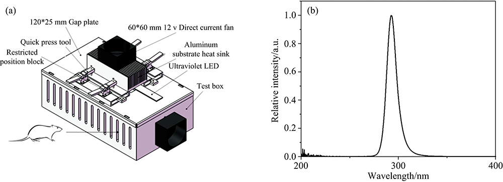 | Fig.1 (a) Schematic of rat irradiation devise with LED module, (b) Relative spectral irradiance of prepared LED modules |
In order to determine the full width and half maximum wavelengths of the LED irradiation device, we used an SPD-200 UV radiometer (Sensing, Hangzhou, China) to measure the spectrum of each LED module. This way, could determine where the peak is 293 nm, and the full width at half maximum is 10 nm (2b), which met our requirements for this study. In order to characterize our rat osteoporosis model, we analyzed the level of 25-hydroxyvitamin and the bone microarchitecture of rats before and 3 months after bilateral ovariectomy. 25(OH)D3 is present at a high concentration in the blood and has a long half-life and is stable nature, so it is a reliable index to reflect the vitamin D levels in the body. As shown in Figure 2. The 25(OH)D3 level in OVX was significantly lower than that of Sham (p< 0.05). The BV/TV, Tb.N and Tb.Th in OVX significantly decreased, while BS/BV and Tb.Sp significantly increased when compared with the Sham (p< 0.05). These results indicated that we had successfully established a rat osteoporosis model.
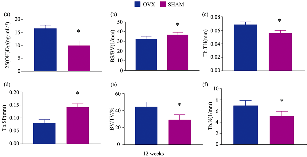 | Fig.2 We had successfully established a rat osteoporosis model (a): 25(OH)D3; (b): Bone surface area/bone volume; (c): Trabecular thickness; (d): Trabecular separation; (e): Bone volume/total volume; (f): Trabecular number; * p< 0.05 |
Serum 25(OH)D3 is a reliable index reflective of the vitamin D levels in the body. As shown in [Figure 3(a)], 4 weeks into the UV LED irradiation treatments, the serum 25(OH)D3 level in the LED irradiation group increased significantly when compared to control rats (p< 0.05, all groups). With continued irradiation treatments through 8 and 12 weeks, the serum 25(OH)D3 level continued to increase and was maintained at a high level (p< 0.05, all groups). However, rats in the OVX6 not exposed to UV LED irradiation had a low level of serum 25(OH)D3 at 4, 8, and 12 weeks, while the Sham6 group maintained a normal level of serum 25(OH)D3 at 4, 8 and 12 weeks.
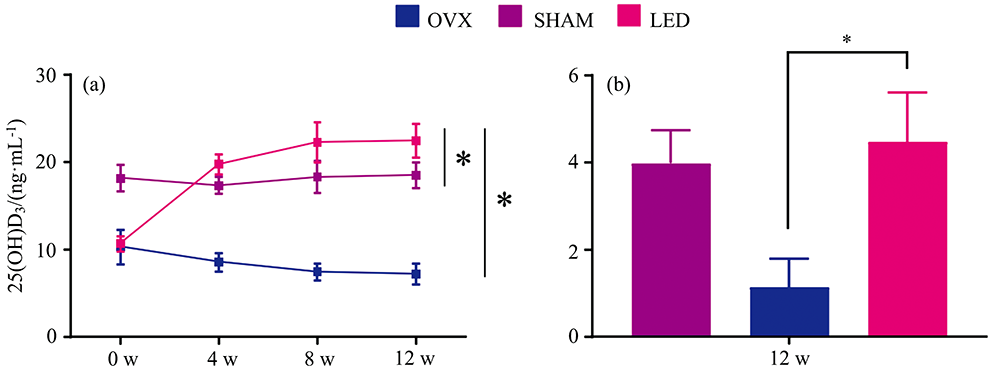 | Fig.3 Effect of irradiation on the serum 25(OH)D3 and expression of vitamin D 25-hydroxylase (a): 25(OH)D3; (b): Relative 25-hydroxylase mRNA; * p< 0.05 |
Serum TRAP is secreted by osteoclasts and is often used as a bone resorption marker. Serum PTH is an important hormone that regulates calcium and phosphorus metabolism in the human body. PTH levels are often observed when the bone resorption rate increases[30]. As shown in Fig.4(a, b), serum PTH and serum TRAP levels decreased significantly following UV irradiation in the LED irradiation group compared to the unirradiated OVX group (p< 0.05). No significant changes were observed in the Sham group's serum PTH and serum TRAP levels before and after the experiment. Phosphorus and calcium are basic substances of bone mineralization and participates in the whole process of bone remodeling. As shown in Figure 4(c) and (f), no significant differences were observed in serum phosphorus and serum calcium between the LED irradiation group, Sham group and OVX group. Serum bone ALP and serum P1NP are markers reflecting bone formation and osteoblast activity[31]. As shown in Figure 4(d) and (e), the serum bone ALP and serum P1NP levels in the LED irradiation group were significantly higher than those of the unirradiated OVX group (p< 0.05). There were no significant changes in serum bone ALP and serum P1NP levels between the OVX group and the Sham group before and after the experiment.
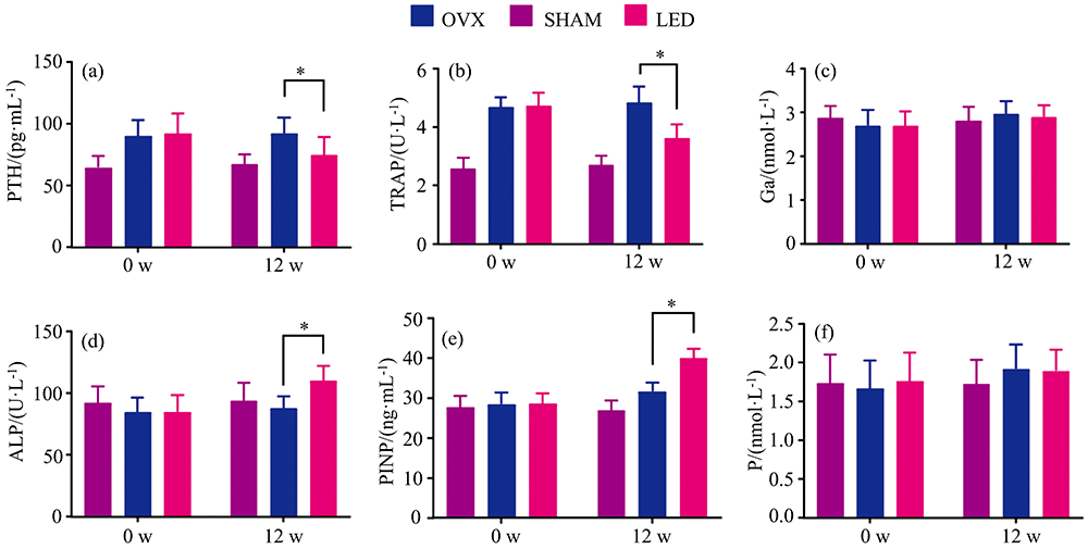 | Fig.4 (a) Parathyroid hormone; (b) Tartrate-resistant acid phosphatase; (c) Serum calcium; (d) Serum phosphorus; (e) Alkaline phosphatase; (f) Procollagen 1N-telopeptide; * p< 0.05 |
Vitamin D 25-hydroxylase can convert vitamin D into 25(OH)D3. In order to detect changes indicative of alterations in 25(OH)D3 metabolism, we studied the mRNA expression level of vitamin D 25-hydroxylase by RT-PCR. Relative expression levels in Sham and LED irradiation groups are expressed with reference to in the OVX group as 1.0. As shown in Figure 3(b), the mRNA level of vitamin D 25-hydroxylase in the LED irradiation group was significantly higher than that in the OVX group. The results showed that vitamin D converted from 7-dehydrocholesterol in UV-irradiated skin is likely sent to the liver, where elevated levels of vitamin D25-hydroxylase could, in turn, convert a large amount of vitamin D to 25(OH)D3, which would explain the significant increase in serum 25(OH)D3 observed in the rats.
Phospho-p53 are proteins whose levels are indicators of the incidence of DNA damage[32]. We therefore, performed IHC analysis of these DNA damage markers in UV LED irradiated tissues from the osteoporosis rats and quantified the cumulative optical density for each marker. The results showed no significant difference in cumulative optical density for either DNA damage marker when comparing the UV LED irradiation group to controls (Figure 5), indicating DNA damage was not induced as a result of treatment.
 | Fig.5 The effects of UV LED irradiation on skin health (a)— (c): IHC with anti-Phospho-p53 antibody for dermis and epidermis (original magnification 400× , bars indicate 100 μm); (d): Cumulative optical density of Phospho-p53 protein detected by IHC |
Barnkob et al.[33] demonstrated that vitamin D could be generated in pig skin when irradiated with a UV LED light source between 292 and 300 nm. However, since the authors chose an in vitro experimental approach, no analysis could be performed to examine the effect of this treatment on bones. Morita et al.[34] found that UV LED irradiation can increase vitamin D production in the body and that short- and medium-term UV irradiation does not change bone microarchitecture. However, the authors note that the selected model is not an osteoporosis model, and the observation period is too short, so limited conclusions can be made regarding the efficacy or safety of this approach for treating osteoporosis. Later studies with this treatment approach were performed in a vitamin D deficiency/accelerated aging mouse model, but serum PTH, calcium, and phosphorus levels, which are closely related to bone metabolism, were not studied. The author of this study indicated that the blood samples collected from the mice were too small to complete these analyses. Therefore, in studying the therapeutic effect of UV LED irradiation on osteoporosis, selecting an appropriate animal model is particularly important. In this study, a rat model of osteoporosis was established through an ovariectomy and administration of a vitamin D-deficient diet, to imitate the characteristics of the disease most common to postmenopausal osteoporosis and osteoporosis caused by diminished vitamin D metabolism in the elderly[35, 36]. We found that administration of short-wave UV LED irradiation at a wavelength of 293 nm can significantly increase the vitamin D level in osteoporotic rats, promote bone formation, and inhibit bone absorption.
Initially, due to the wide wavelength spectrum, large size, and the high price of fluorescent tubes capable of UV radiation, there had been no breakthroughs in this area regarding osteoporosis treatment. Now, with LED technology's continuous development and Al components' adjustment in AlGaN materials, various narrow spectra can be arbitrarily adjusted and delivered using UV LED irradiation devices. Furthermore, with the transition from a traditional semiconductor to a flexible semiconductor, the construction of wearable LED medical devices is becoming possible[37, 38]. Compared with other UVB light sources, LED sources produce the least radiant heat, enhancing the comfort of wearable UV LED devices[39].
Although UV radiation can improve bone mineral density and bone mineral content in rats, this study has some limitations. First, we only studied the effect of a single UV LED irradiation dose on bone metabolism, bone microarchitecture, and skin health in rats. The potential risk of skin inflammation may be higher with higher doses, and too low a dose may reduce this approach's therapeutic benefit in treating osteoporosis. Therefore, it would be worthwhile to study the dose-effect relationship of the effects of UV LED irradiation on bone microarchitecture, bone metabolism and skin in osteoporotic rats in a future study. Finding an optimal dose range will be of great significance for developing LED treatment equipment for osteoporosis in patients[40, 41]. Second, this experiment did not study the effect of UV LED irradiation on mood and immune regulation in rats. Therefore, the optimization of UV LED irradiation wavelengths, time of exposure, and dose are necessary for the future application of UV LED irradiation in the treatment of osteoporosis and the development of wearable UV LED devices[42, 43].
In summary, this study demonstrates that UV LED irradiation can significantly increase the blood 25(OH)D3 level in osteoporotic rats, suppress bone resorption, enhance bone formation, and improve bone microarchitecture. The data provide useful information for future studies, and the application of UV LED irradiation for the treatment of osteoporosis and the development of wearable UV LED medical devices. With the increases in life expectancy in many developed countries, there is a greater incidence of chronic diseases such as osteoporosis[44, 45]. Therefore, it will be of great economic and social importance to develop safe, effective, low-cost, portable and wearable non-invasive phototherapy equipment for the treatment of osteoporosis.
Conflict of interest statement
The authors declare no conflict of interest in any form concerning this article.
| [1] |
|
| [2] |
|
| [3] |
|
| [4] |
|
| [5] |
|
| [6] |
|
| [7] |
|
| [8] |
|
| [9] |
|
| [10] |
|
| [11] |
|
| [12] |
|
| [13] |
|
| [14] |
|
| [15] |
|
| [16] |
|
| [17] |
|
| [18] |
|
| [19] |
|
| [20] |
|
| [21] |
|
| [22] |
|
| [23] |
|
| [24] |
|
| [25] |
|
| [26] |
|
| [27] |
|
| [28] |
|
| [29] |
|
| [30] |
|
| [31] |
|
| [32] |
|
| [33] |
|
| [34] |
|
| [35] |
|
| [36] |
|
| [37] |
|
| [38] |
|
| [39] |
|
| [40] |
|
| [41] |
|
| [42] |
|
| [43] |
|
| [44] |
|
| [45] |
|