


一种用于水中次氯酸根检测的高选择性裸眼比色探针
[余青1  , 陈晓丽
, 陈晓丽2 , 张奇龙1, 3, * , 刘华3 , 杨先炯3 , 徐红3 , 黄亚励3, * , 冯星4, * , REDSHAW Carl5 ]
, 陈晓丽, 张奇龙, 刘华, 冯星, REDSHAW Carl|
|


实时检测和监测水中的次氯酸根离子(ClO-)是极富挑战性的研究工作。 报道了一种光学性能优异、 “裸眼”可分辨的比色型探针分子(PAH)。 首先利用高分辨质谱, 核磁共振氢谱和核磁共振碳谱等方法对目标探针分子(PAH)的结构进行表征。 随后, 利用紫外-可见吸收光谱考察了不同pH缓冲溶液条件下探针PAH与次氯酸根离子的相互作用。 结果显示, 水溶性的探针分子PAH在pH值为2.0~5.0的磷酸盐缓冲液中为黄色溶液, 其最大吸收峰在424 nm处; 在pH值为6.0~12.0的磷酸盐缓冲液中PAH为紫色溶液, 最大吸收峰在532 nm处; 在不同pH缓冲液体系中分别加入次氯酸根离子, 肉眼可观察PAH溶液颜色褪去, 紫外-可见吸收光谱显示在424 nm处的特征吸收峰逐渐降低并在532 nm处出现新的吸收峰, 溶液颜色从黄色到紫色然后到无色, 特征峰明显消失。 进一步优化了实验条件, 发现在pH 5.0的磷酸盐缓冲液中, 探针分子PAH对ClO-离子具有特定的选择性和灵敏度, 并且具有较低检出限等优点; 在优化的条件下, 探究了常见的金属离子、 阴离子等共存条件下, 对探针分子PAH检测次氯酸根离子的干扰影响。 实验发现, 常见的金属离子(Li+, Co2+, Cr3+, K+, Cd2+, Pb2+, Ca2+, Hg2+, Ba2+, Cu2+, Mg2+, Ni2+, Zn2+, Al3+和Fe3+), 阴离子($NO_{2}^{-}$, I-, AcO-, $ClO_{4}^{-}$, $SO_{4}^{2-}$, CN-, Br-, $CO_{3}^{2-}$和F-), 活性氧(ROO·, ·OH, H2O2, · $O_{2}^{-}$,tBuOOH,tBuO·和1O2), 和活性氮(ONOO-和NO·)等33种物质对探针分子检测ClO-离子的干扰较小。 同时, 探针PAH可以定量检测次氯酸根离子( y=1.586 78-0.524 51 x, R2=0.998 52), 检出限为5.39 μmol·L-1。 此外, 对水体系(84消毒剂和自来水)中的次氯酸根离子浓度进行分析, 三次平行试验测得自来水中次氯酸根离子的平均浓度为7.96 μmol·L-1, 平均加标回收率高, 表明探针PAH还可用于定量检测实际水体系中的次氯酸根离子。
, CHEN Xiao-li, ZHANG Qi-long, LIU Hua, FENG Xing, REDSHAW CarlBiography: YU Qing, (1992—), postgraduate in School of Public Health, the Key Laboratory of Environmental Pollution Monitoring and Disease Control, Ministry of Education, Guizhou Medical University e-mail: 1432507000@qq.com;CHEN Xiao-li, (1996—), graduate in School of Clinical Medicine, Guizhou Medical University e-mail: 2422691291@qq.com
The real-time detection and monitoring of hypochlorites (ClO-) in water is highly challenging. An excellent colorimetric “naked-eye” probe photoacid (PAH) was synthesized. PAH was comfirmed using High-Resolution Mass Spectrometry (HRMS),1H NMR and13C NMR. The interaction between PAH and ClO- was investigated via UV-Vis absorption spectrophotometry under different pH conditions. The results indicated that PAH was completely soluble in water, PAH displayed a yellow solution in a phosphate buffer with a pH of 2.0 to 5.0, and the maximum absorption peak was at 424 nm. PAH displayed a purple solution in a phosphate buffer with a pH of 6.0~12.0, and the maximum absorption peak was at 532 nm. After adding ClO- to different pH systems, PAH discoloration and the UV-Vis absorption peak disappeared. The probe PAH exhibited specific selectivity and sensitivity for ClO- detection with a low detection limit in the pH 5.0 phosphate buffer. After PAH reacted with ClO-, the absorption peak of the probe at 424 nm gradually decreased, and a new absorption peak appeared at 532 nm. The probe displayed a vivid color-tunable process from yellow to purple then to colorless with a fast response time for ClO- detection. However, other common 33 substances such as metal ions(Li+, Co2+, Cr3+, K+, Cd2+, Pb2+, Ca2+, Hg2+, Ba2+, Cu2+, Mg2+, Ni2+, Zn2+, Al3+ and Fe3+), anions ($NO_{2}^{-}$, I-, AcO-, $ClO_{4}^{-}$, $SO_{4}^{2-}$, CN-, Br-, $CO_{3}^{2-}$ and F-), reactive oxygen species (ROO·, ·OH, H2O2, · $O_{2}^{-}$, tBuOOH, tBuO· and1O2) and reactive nitrogen species (ONOO- and NO·), did not cause changes in the color of the probe solution and the UV-Vis absorption spectrum. The above species had only a limited effect on detecting the ClO- anion. When they coexisted with ClO-, the probe also showed a similar solution color change, and the absorption peak at 424 nm disappeared. Meanwhile, the probe PAH could quantitatively detect the content of ClO- with a detection limit of 5.39 μmol·L-1 ( y=1.586 78-0.524 51 x, R2=0.998 52). Furthermore, ClO- concentration in the water system (84 disinfectant and tap water) was analyzed. The average concentration of ClO- ion in the tap water measured by three parallel tests was 7.96 μmol·L-1 with high recoveries rate. It showed that PAH could also be utilized to detect ClO- quantitatively in real water systems.
Hypochlorous acid (HClO) or hypochlorite (ClO-), as typical reactive oxygen species (ROS), play several fundamental roles in the human body and are biologically produced by the reaction of chloride ions (Cl-) and hydrogen peroxide (H2O2) via catalysis of myeloperoxidase (MPO) in the immune cell[1]. Moreover, an appropriate amount of ClO- can protecting the immune system against the invasion of pathogens. Nevertheless, excess production of ClO- may lead to ageing and an increased risk of cell membrane damage[2], Alzheimer’ s disease[3] and cardiovascular disease[4].
Hypochlorite is ubiquiuous in daily human life and appears in many applications, such as the sanitization of tap water and swimming pools. Traditional methods for the detection of ClO- include iodine reduction titration, electrochemical methods[5], chemiluminescence methods[6] and ion chromatography[7] as well as spectroscopic (colorimetry and fluorescence) detection. Commonly, HClO/ClO- concentration in the standards for drinking water quality in China ranges from 0.05 to 4 mg· L-1[8]. Theoretically, the detection mechanism of HClO is involved in common oxidation reactions[9], cleavage reactions[10]. However, the hydrophobic nature of the probe has limited their practical application environment. Water-solubility is a crucial criterion for the practical use of a probe; and the development of water-solubility colorimetric probes with a specific response to ClO- and HClO in a water environment still remain challenging. There are few reports on the detection of ClO- and HClO in a water environment by use of colorimetric probes. Therefore, it is important to design and synthesize hypochlorite colorimetric probes with good water-solubility, high selectivity and sensitivity. More importantly, this type of probe with excellent characteristics can be used for rapid and selective detection of HClO/ClO- in water environments.
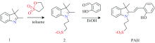
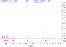
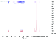
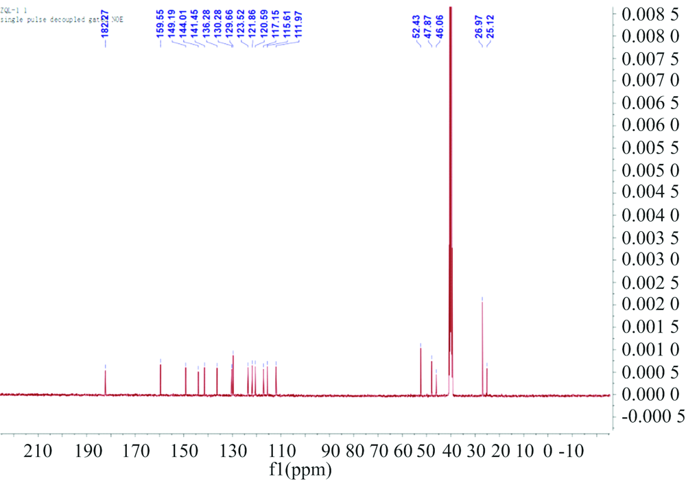
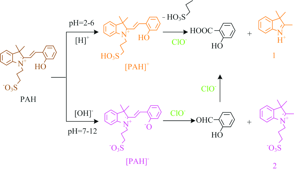
In this paper, for portable purposes, a simple-to-use and naked-eye diagnostic tool were explored for ClO- detection in an aqueous solution with the advantage of a rapid response, high sensitivity and a low detect limitation. Herein, we present an example of a colorimetric “ naked-eye” probe photoacid (PAH) for ClO- detection with high selectivity in aqueous solution, which was designed and synthesized via a Knoevenagel reaction between indolinium and salicylic aldehyde in good yield[11] (Scheme 1). The structures of the synthesized compounds were fully characterized by 1H/13C NMR spectroscopy and High-Resolution Mass Spectrometry (HRMS); the spectra are shown in the Supplementary Information (Fig.S1—S2).
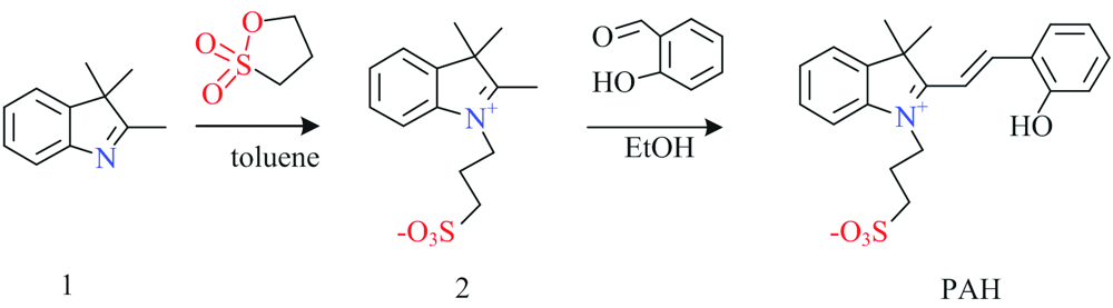 | Scheme 1 The synthetic route to probe PAH |
 | Fig.S1 1H NMR spectrum of PAH (400 MHz, d6-DMSO) |
 | Fig.S2 13C NMR spectrum of PAH (d6-DMSO) |
All reagents and solvents were of analytical grade and were used without further purification. Ultrapure water was used throughout the experiments. The solutions of metal ions were prepared from their nitrates, and the solutions of anions were prepared from their sodium salts. The UV-Vis absorption spectra were determined at room temperature on a Shimadzu UV-2600 spectrophotometer in a 1 cm quartz cell. The pH values were determined with a model pHS-25 pH meter. High-resolution mass spectrometry (HRMS) were performed in a micro TOF-QⅡ mass spectrometer (USA). 1H/13CNMR (400 MHz) spectra was recorded on a Bruker Advance 400 spectrometer(Germany), with DMSO-d6 used as a solvent and tetramethylsilane (TMS) as an internal standard.
1.2.1 Synthesis of probe PAH
Probe PAH was synthesized according to the reported procedures[12]. A mixture of 2, 3, 3-trimethylindolenine 1 (1.65 g, 0.01 mmol) and 1, 2-oxathiolane 2, 2-dioxide (1.26 g, 0.01 mmol) in toluene and stirred at 90 ℃ for 4 h under N2. The purple solid was collected by filtration, washed with cold ethylether, and dried in vacuo to afford 2, 3, 3-trimethyl-1-(3-sulfonatepropyl)-3H-indolium 2 (2.61 g, 89% yield). Without further purification, the synthesized compound 2 (100 mg, 0.36 mmol) and 2-hydroxybenzaldehyde (48 mg, 0.39 mmol) were added into anhydrous ethanol (2 mL). The mixture was allowed to reflux overnight. The orange solid was obtained by filtration (110 mg, 80% yield); 1H NMR (400 MHz, d6-DMSO): δ 8.6~8.48 (m, 1H), 8.24 (d, J=7.9 Hz, 1H), 7.99 (d, J=8.2 Hz, 1H), 7.90~7.78 (m, 2H), 7.65~7.53 (m, 2H), 7.44 (t, J=7.7 Hz, 1H), 7.04~6.89 (m, 2H), 4.90~4.66 (m, 2H), 2.62 (t, J=6.4 Hz, 2H), 2.22~2.05 (m, 2H), 1.84~1.62 (m, 6H); 13C NMR (100 MHz, d6-DMSO) δ 182.27 (s), 159.55 (s), 149.19 (s), 144.01 (s), 141.45 (s), 136.28 (s), 130.28 (s), 129.66 (s), 123.52 (s), 121.86 (s), 120.59 (s), 117.15 (s), 115.61 (s), 111.97 (s), 52.43 (s), 47.87 (s), 46.06 (s), 26.97 (s), 25.12 (s). HRMS: m/z [M+Na]+=408.126; Calcd: 407.120. The 1H/13C NMR data is consisting with the previously reported data[12].
1.2.2 Preparation of solutions
Phosphate buffer saline (PBS) with different pH from 2.0 to 12.0 was prepared from disodium hydrogen phosphate, dihydrogen phosphate and sodium chloride in a certain proportion with ultrapure water[13]. The 0.5 mmol· L-1 stock solution of probe PAH was prepared in an aqueous solution of PBS (0.01 mol· L-1, pH 5.0). The 0.01 mol· L-1 stock solutions of the metal ions and various anions were dissolved in an aqueous solution of PBS (0.01 mol· L-1, pH 5.0). The 0.01 mol· L-1 stock solutions of reduced vitamin C (Vc) and glutathione (GSH) were prepared directly from the solids in aqueous solution of PBS (0.01 mol· L-1, pH 5.0). Hypochlorites were derived from sodium hypochlorite. The stock solution of ClO- was prepared with 0.01 mol· L-1 sodium hydroxide solution and standardized at 292 nm using an extinction coefficient 350 M-1· cm-1 at pH 12.0[14], then diluting the stock solution of ClO- to 8 mmol· L-1. The 0.01 mol· L-1 stock solution of other reactive oxygen species (ROS) and reactive nitrogen species (RNS) were prepared according to the literature[15].
1.2.3 General procedure for analysis
Before conducting the spectroscopic measurements, the corresponding solutions of probe PAH and the reactive species (metal ions, anions, ROS and RNS) were freshly prepared. For UV-Vis selective experiments, test solutions were prepared by placing 0.6 mL of the probe PAH solution (0.5 mmol· L-1) and 0.12 mL of reactive species solutions in the absence and presence of 0.12 mL of ClO- solutions (8 mmol· L-1) into a 3 mL cuvette, and then diluting the solution to 3 mL with PBS (0.01 mol· L-1, pH 5.0). For UV-Vis titrations, 0.6 mL of the probe PAH solution and different amounts of ClO- was added into a 3 mL cuvette. These solutions were diluted with PBS (0.01 mol· L-1, pH 5.0) to 3 mL and mixed, then the spectra of these solutions were immediately recorded through the UV-Vis method.
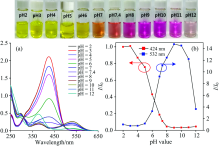
Previous reports indicated that PAH would undergo decomposition depending on the pH of the aqueous solution[16]. Thus, the pH-dependent stability of PAH was investigated in different buffered solutions from pH 2.0 to 12.0. As shown in Fig.1, the probe in pH 2 solution exhibited a yellow color and a well-resolved absorption band range from 300~500 nm with a maximum absorption peak at 424 nm, as the pH increased from 2.0 to 5.0, the absorption peak at 424 nm gradually decreased, which may be due to protonation of the terminal
 | Fig.1 The change of UV-Vis spectra (a) and line graph (b) of probe PAH (0.1 mmol· L-1) in the range from pH 2.0~12.0 using PBS as buffer solution; The color change of photography of probe PAH with the pH value increases under natural light (inset) |
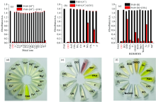
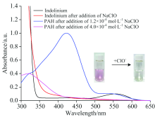
A colorimetric probe’ s high selectivity and sensitivity are key parameters for real applications in tap water monitoring and in vivo studies. Thus, the UV-Vis response of PAH toward metal ions and anions, reactive oxygen species (ROS) and reactive nitrogen species (RNS) was evaluated in pH 5.0 buffered solution as presented in Fig.2 and in Fig.S3. Clearly, except for ClO-, the PAH solution (0.1 mmol· L-1) still displays a yellow color, and the corresponding UV-Vis spectra indicated that the absorption behavior exhibited slight changes after the addition of 4 equiv. The various extra species, such as metal ions and anions, reactive oxygen species (ROS) and reactive nitrogen species (RNS). Furthermore, by adding the same concentration of ClO- into the solution containing the PAH and the metal ion species, the absorption peak at 424 nm disappeared and the solution color changed from yellow to colorless [Figure 2 (a) and (d)]. The results indicated that the PAH exhibited a highly selective response toward ClO- with great anti-interference ability to other species. Furthermore, when the ClO- adding into the mixture of PHA and various anions, the UV-Vis spectra show only slight changes excepted the reductive anions, such as I-, ONOO-,
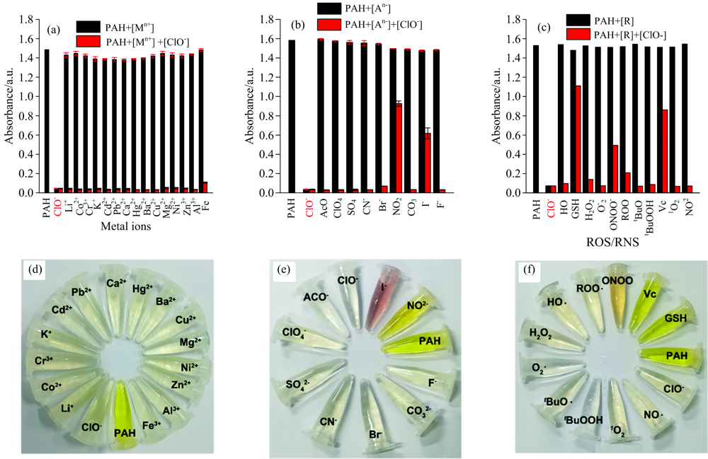 | Fig.2 Absorption intensity (a—c) at 424 nm of probe PAH (0.1 mmol· L-1) with addition of different species (0.4 mmol· L-1) in the absence/presence of ClO- (0.4 mmol· L-1) in PBS solution (0.01 mol· L-1, pH 5.0), Error bar=RSD (n=3); Visible color (d—f) of probe PAH toward ClO- (0.4 mmol· L-1) in different species under sunlight |
 | Fig.S3 UV-Vis spectra of PAH (0.1 mmol· L-1) with addition of nitrate salts of Li+, Co2+, Cr3+, K+, Cd2+, Pb2+, Ca2+, Hg2+, Ba2+, Cu2+, Mg2+, Ni2+, Zn2+, Al3+ and Fe3+(0.4 mmol· L-1), sodium salts of |
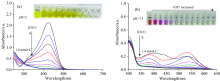
Further, the effect of I-, ONOO-,
 | Fig.3 UV-Vis absorption spectra (b) of PAH (0.1 mmol· L-1) in the presence of I- toward various concentration of ClO- in PBS buffer solution (0.01 mol· L-1, pH 5.0); The change of visible color (a) of probe PAH toward ClO- under sunlight |
 | Fig.S4 UV-Vis absorption spectra (a—d) of PAH (0.1 mmol· L-1) with the addition of |
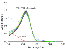
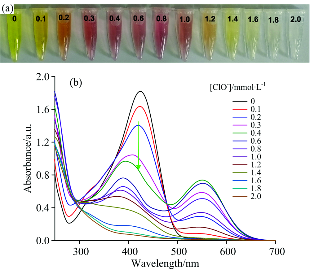
The recognition ability of probe PAH toward ClO- was measured using UV-Vis titrations in buffered aqueous solution (PBS, 0.01 mol· L-1, pH 5.0). As shown in Fig.4, with increasing ClO- concentrations, the absorption peak of probe PAH at 424 nm gradually decreased and the short absorption wavelength centering around 296 nm gradually increased, accompanied by a change in the color of the solution from yellow to colorless. Moreover, a well-defined isosbestic point was observed at 315 nm, indicating the formation of a single new species. In addition, the UV-Vis titration experiments between the corresponding absorbance values at 424 nm and ClO- concentrations exhibited an excellent linear correlation with a high coefficient (y=1.586 78-0.524 51x, R2=0.998 52) in the range of 0~0.28 mmol· L-1 [Fig.4(c)].Thus, the detection limit of probe PAH for ClO- detection was determined to be 5.39 μmol· L-1 according to the IUPAC definition (CDL=3Sb/m, Sb is the standard deviation of the blank samples, m is the slope of the linear equation) from 10 blank solutions[18]. These results illustrated that probe PAH has the excellent capability for the qualitative and quantitative determination of ClO- with high sensitivity in total aqueous solutions.
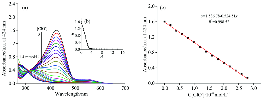 | Fig.4 UV-Vis spectra (a) of probe PAH (0.1 mmol· L-1), changing of the absorption intensity (b) at 424 nm of probe PAH upon addition of increasing concentrations (0~1.4 mmol· L-1) of ClO- in PBS (0.01 mol· L-1, pH 5.0) and linearity of absorption intensity (c) of probe PAH with the addition of ClO- from 0~0.28 mmol· L-1. Error bar=RSD (n=3) |
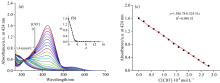
It is noteworthy that on the addition of a small amount of ClO- to the solution of probe PAH, a new absorption band appeared in the range from 500~570 nm, while following an increase in the concentration of ClO-, the absorption gradually disappeared again. We hypothesized that the new long absorption band originated from the degraded compound of indolinium, which strongly agrees with our experiment results (Fig.S5).
 | Fig.S5 UV-Vis absorption spectra of indolinium in the absence/presence of ClO- in aqueous solution and UV-Vis absorption spectra of probe PAH (0.1 mmol· L-1) in the presence of ClO- (0.12 mmol· L-1 or 0.4 mmol· L-1) in PBS (0.01 mol· L-1, pH 5.0); Photography of indolinium in the absence/presence of ClO- (inset) |
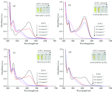
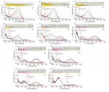
The pH value of the environment is a crucial parameter that can affect the selectivity, sensitivity, and detection limit of a probe[19]. Carefully, the absorption behavior of probe PAH toward ClO- was investigated over the relevant pH range from 2.0 to 12.0, respectively (Fig.5 and Fig.S6). The maximum absorption peak of probe PAH is at 424 nm within a pH range of 2.0~5.0 with a yellow color solution, but when treated with ClO-, the solution color dramatically changed from yellow to colorless, and the maximum absorption peak of 424 nm disappeared. On the other hand, the neutralized PAH at pH> 6.0, displayed two absorption peaks at 424 and 550 nm, respectively. However, when treated with ClO- from 0 to 1.4 mmol· L-1, both absorption peaks gradually decreased, and even disappeared, and the solution containing PAH turned from purple to colorless. These results indicated that the PAH is a highly sensitive, “ naked-eye” colorimetric sensor for ClO- detection in aquatic environments. More importantly, the presence of metal ions and anions, ROS and RNS only provide a limited ClO- detection disruption. Thus the excellent probe PAH can be applied for detecting the ClO- in natural water samples with significantly more complex compositions.
 | Fig.5 UV-Vis spectra of probe PAH (0.1 mmol· L-1) with the addition of ClO- (0~1.4 mmol· L-1) in PBS (0.01 mol· L-1) in pH 2.0 (a), pH 7.4 (b), respectively; The photography change of visible color of probe PAH toward ClO- under sunlight (inset) |
 | Fig.S6 UV-Vis spectra of probe PAH (0.1 mmol· L-1) with the addition of ClO- (0~1.4 mmol· L-1) in PBS (0.01 mol· L-1) in pH 3.0 (a), pH 4.0 (b), pH 5.0 (c), pH 6.0 (d), pH 7.0 (e), pH 8.0 (f), pH 9.0 (g), pH 10.0 (h), pH 11.0 (i), pH 12.0 (j), respectively; The photography change of visible color of probe PAH toward ClO- under sunlight (inset) |
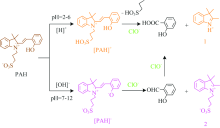
The pH-dependence of the UV-Vis spectra showed that the probe PAH exhibited color-tunable properties from yellow to purple then to pink. There were two pathways to the hydrolysis of PAH in acidic and alkaline environment. In acidic environments (pH 2.0~5.0), the H+ would attack the
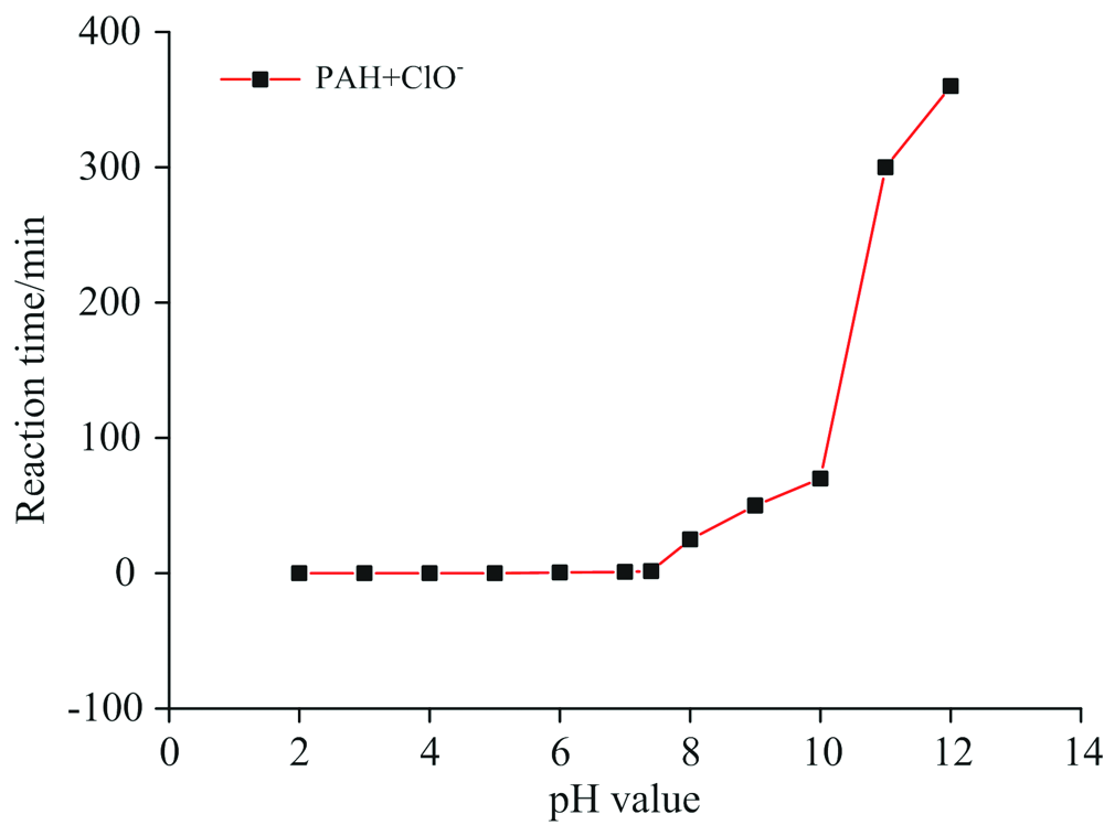 | Fig.S7 Comparison of the interaction time between PAH (0.1 mmol· L-1) and ClO- (0.8 mmol· L-1) in aqueous solution of PBS (0.01 mol· L-1, pH 2.0~12.0) |
 | Scheme 2 The proposed recognition mechanism of probe PAH toward ClO- |
Firstly, in the presence of ClO-, oxidation cleavage of the C=C double of PAH would afford the 2-hydroxybenzoic acid/salicylaldehyde and indolinium derivative depending on the pH conditions. In the pH range from 7.0 to 12.0, when the [PAH]- encountered ClO-, it would be oxidized to 2 and salicylaldehyde, while at pH 2.0~6.0, the [PAH]+ can be oxidized to indolinium fragment 1 with 2-hydroxybenzoic acid. The detailed information of the molecular structures was supported by mass spectrometry and is listed in Fig.S8—Fig.S10.
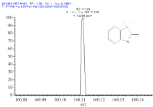
 | Fig.S8 HRMS of intermediate 1 The peak (m/z) at 160.111 89 corresponds to [M+H]+ ion (Calcd: 159.10) |
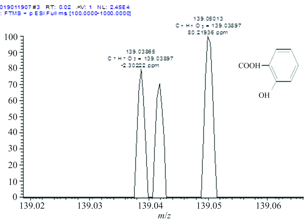 | Fig.S9 HRMS of 2-hydroxybenzoic acid The peak (m/z) at 139.038 97 corresponds to [M+H]+ ion (Calcd: 138.03) |
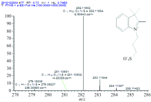
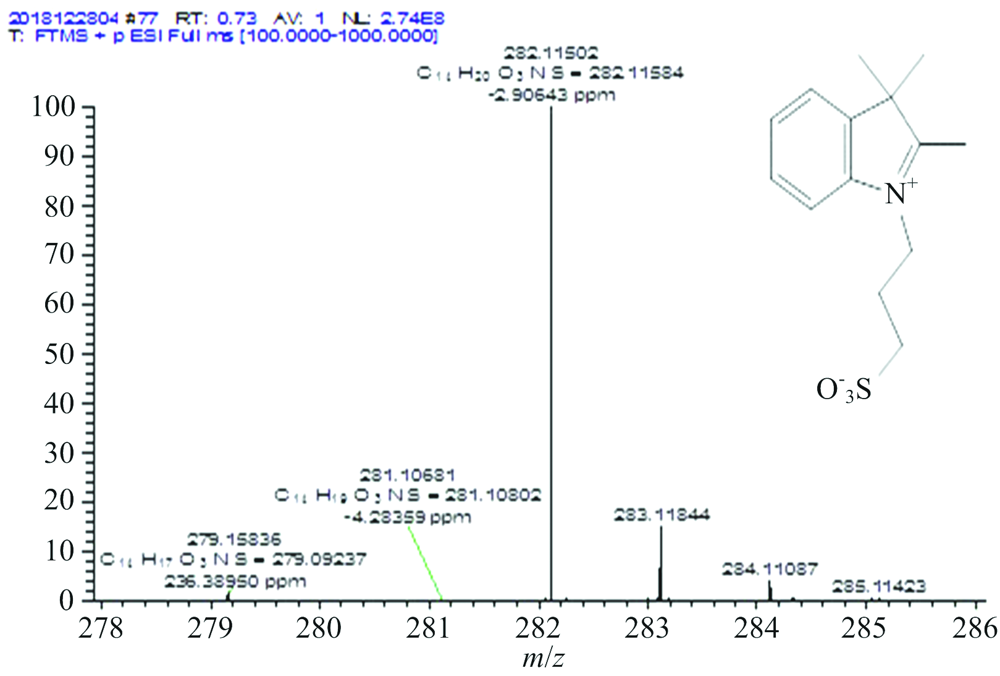 | Fig.S10 HRMS of intermediate 2 The peak (m/z) at 282.115 84 corresponds to [M+H]+ ion (Calcd: 281.11) |
On titration of hypochlorite under acidic conditions, HRMS data manifested the peaks at m/z=160.111 89 and 139.038 97, corresponding to the intermediates 1 and 2-hydroxybenzoic acid; while under basic conditions, HRMS gives result for m/z=123.044 06 and 282.115 84 consistent with intermediate 2 and salicylaldehyde, respectively.
Hypochlorite is widely used in our daily life. Thus, the probe PAH had been applied to analyse the content of ClO- in real samples (such as 84 disinfectant and tap water). The real sample was filtered for the removal of insoluble species. In a PBS (0.01 mol· L-1, pH 5.0) solution of 3 mL containing the probe PAH (0.1 mmol· L-1), the sample of 84 disinfectants was added with different volumes of 2.0, 4.0, 6.0 μL, respectively. The calculated concentration of ClO-of this 84 disinfectant sample is (151± 6.68) mmol· L-1 (Table 1), According to the linear regression equation of UV-Vis titration (y=1.586 78-0.524 51x, R2=0.998 52). Further, the content of ClO- in tap water and purified water were also measured under the same conditions, and the calculated concentration of ClO- is 7.96 μmol· L-1 for the tap water sample (Table 1). Except for one sample, the percentage recoveries were in the range of 93%~103%. These results indicated that the probe PAH could potentially be used for the quantitative detection of ClO- concentrations in real water samples.
| Table 1 Determination of ClO- concentrations in real samples |
In conclusion, based on the oxidative activity of ClO-, the PAH can be utilized as a highly selective colorimetric naked-eye probe for ClO- detection via oxidative cleavage of itself in absolute aqueous solution, which leads to a tunable color process from yellow to purple then to colorless. The specificity of probe PAH can work in water environments, and coexisting ions only show a slight effect on ClO- detection. More importantly, acidic solid conditions accelerate the response time. Moreover, PAH exhibited a fast response time and naked eye detection toward ClO- at various pH values with highly-sensitive detection at pH 5.0. We have also successfully applied the probe to detect ClO- in real water samples. This research will open up new avenues for developing novel “ naked-eye” probes for the detection of environmental pollutants in an absolute aqueous system.
| [1] |
|
| [2] |
|
| [3] |
|
| [4] |
|
| [5] |
|
| [6] |
|
| [7] |
|
| [8] |
|
| [9] |
|
| [10] |
|
| [11] |
|
| [12] |
|
| [13] |
|
| [14] |
|
| [15] |
|
| [16] |
|
| [17] |
|
| [18] |
|
| [19] |
|