
SiNWs:Tb3+的光学稳定性及Si纳米线对其发光特性的影响
[杨瑞臣1  , 耿小丕
, 耿小丕1 , 范志东1, * , 李旭2 , 马蕾3 , 高永慧1 , 付莹1 , 王欣欣4 ]
, 耿小丕, 李旭|
|


作者简介: 杨瑞臣, 1978年生, 承德石油高等专科学校数理部讲师 e-mail: huoxiangzhengqishui@126.com
荧光纳米材料不但具备纳米材料的优势, 同时还具有优异的光学性质, 被广泛应用于荧光标记、 离子识别、 荧光免疫分析、 光学成像和医学诊断等方面。 因此, 荧光纳米材料的制备、 结构分析和荧光特性等方面的研究备受人们的关注。 为了获得发光强度大、 荧光量子效率高和制备过程可控的Si基荧光纳米材料, 实验进一步研究了Si纳米线对样品发光特性的影响和样品的光学稳定性。 首先, 基于固-液-固生长机制, 在反应温度为1 100 ℃、 N2气流量为1 500 sccm、 生长时间为15~60 min等工艺条件下, 分别以“抛光”和“金字塔”织构表面的单晶Si(100)为衬底, 生长出不同长度和分布的Si纳米线; 以Au或Au-Al合金膜层作为金属催化剂, 生长出密度分别约为108和1010 cm-2的Si纳米线; 然后, 利用L4514自动控温管式加热炉, 基于高温固相法, 在温度为1 100 ℃、 掺杂时间为60 min和N2气流量为1 000 sccm等工艺条件下, 以高纯Tb4O7(99.99%)粉末为稀土掺杂剂对不同Si纳米线衬底进行稀土掺杂, 制备一系列的荧光纳米材料SiNWs:Tb3+样品; 室温下利用Hitachi F-4600型荧光分光光度计, 固定激发光波长为243 nm、 激发光狭缝为2.5 nm、 发射光狭缝为2.5 nm、 扫描波长范围为450~650 nm、 光电倍增管(photomultiplier lube, PMT)电压为600 V等参数下, 测量了不同样品的光致发光特性; 最后, 实验测试了该荧光纳米材料的光学稳定性, 如时间(0~30 d)、 温度(300~500 K)、 酸碱(pH 1和11)、 抗光漂白(0~120 min)等稳定性以及水溶性和分散性。 结果显示, 在衬底为“金字塔”织构表面上、 生长时间为30 min、 以Au为金属催化剂等条件下制备的Si纳米线为Tb3+掺杂衬底时, SiNWs:Tb3+的绿光发射强度较大, 其发光强峰值位于554 nm, 属于能级5 D4→7 F5的跃迁, 另外在波长为494, 593和628 nm出现了三条发光谱带, 它们分别属于能级5 D4→7 F6,5 D4→7 F4和5 D4→7 F3的跃迁。 另外, 样品展示出了优异的时间、 温度、 酸碱和抗光漂白等光学稳定性, 同时还具有良好的水溶性和分散性。 如温度升高到500 K时, 光发射强度仅降低了约8.9%左右; 抗光漂白能力较强, 用波长为365 nm、 功率为450 W的紫外光源照射120 min, 样品的绿光发射强度无衰减; 酸、 碱稳定性好, 在pH 1的强酸(HCl)溶液中120 min未见衰减, 在pH 11的强碱(NaOH)溶液中15 min内衰减较小, 随后发光强度出现了缓慢下降的趋势; 当60 min后, 样品的发光强度变得极其微弱。 分析认为, 在SiNWs:Tb3+表面有一层SiO2包覆层, 而NaOH溶液容易和SiO2发生化学反应, 随着时间延长SiO2层被破坏, 故样品发光强度降低; 样品溶于水中放置30 d未见沉淀物, 发光亮度均匀且分散性较好。 在研究了制备温度、 气体流量和掺杂时间等工艺条件之后, 深入研究了Si纳米线自身变化对Tb3+绿光发射的影响。 该材料展示出了良好的光学稳定性、 水溶性和分散性, 使其作为荧光标记物具有一定的应用价值。
, GENG Xiao-pei, LI XuFluorescent nanomaterials not only have the advantages of nanomaterials, but also have excellent optical properties. They are widely used in fluorescent labels, ion recognition, fluorescence immunoassays, optical imaging and medical diagnostics. Therefore, researches on the preparation, structure analysis and fluorescence characteristics of fluorescent nanomaterials have received much attention. In order to obtain Si-based fluorescent nanomaterial with high luminous intensity, high fluorescent quantum efficiency and better controlling of preparation process, the effects of Si nanowires on the luminescence properties and the optical stabilities of the samples were investigated. First, based on the solid-liquid-solid growth mechanism, Si nanowires were grown under the conditions of reaction temperature of 1 100 ℃, N2 gas flow rate of 1 500 sccm and growth time of 15~60 min. The Si nanowires with different lengths and distributions were grown on single-crystal Si(100) substrates with “polishing” and “pyramid” texture surfaces respectively. And the Si nanowires with density of about 108 and 1010 cm-2 were grown with Au or Au-Al alloy film as metal catalyst. Then, a series of fluorescent nanometers SiNWs:Tb3+ samples were prepared using L4514 automatic temperature control tube heating furnace based on high-temperature solid-state method under the temperature 1 100 ℃, doping time 60 min, N2 gas flow rate 1 000 sccm, different Si nanowire substrates and high-purity Tb4O7 (99.99%) powder as doping agent. Next, at room temperature, the photoluminescence characteristics of different samples were measured by the Hitachi F-4600 fluorescence spectrophotometer with the fixed excitation light wavelength of 243 nm, the excitation light slit of 2.5 nm, the emission light slit of 2.5 nm, the scanning wavelength range of 450~650 nm and the PMT voltage of 600 V. Finally, the optical stabilities of the fluorescent nanomaterial was experimentally tested, such as time stability (0~30 d), temperature stability (300~500 K), acid and alkali stability (pH 1 and pH 11), anti-photobleaching stability (0~120 min), etc. Besides the water solubility and the dispersibility were tested. The results showed that SiNWs:Tb3+ produced strong green light emission with Si nanowires substrate which was prepared with the growth time 30 min, the “pyramid” texture surface and Au as the metal catalyst. The peak of green emission was 554 nm, which belongs to energy level transition5 D4→7 F5. At the same time, three bands appeared at wavelengths of 494, 593 and 628 nm, which belong to the energy levels transitions5 D4→7 F6,5 D4→7 F4 and5 D4→7 F3, respectively. In addition, the sample was shown to have excellent optical stabilities such as time, temperature, acid-base, anti-photobleaching, good water solubility and dispersibility. When the temperature is increased to 500 K, the light emission intensity is reduced by only about 8.9%. The green light luminescence intensity of the sample is not attenuated when irradiated with an UV light source with a wavelength of 365 nm and a power of 450 W for 120 min. In the strong acid solution of pH=1, no attenuation was found in 120 min and the attenuation was less in 15 min in the strong alkaline solution of pH 11. Subsequently, the luminous intensity showed a slow decline, but the luminous intensity of the sample became extremely weak after 60 min. The analysis showed that there is a layer of SiO2 coating on the surface of SiNWs:Tb3+, and NaOH solution easily reacts with SiO2. As time increases, the SiO2 layer is destroyed, so the luminescence intensity of the sample decreases. There was no deposit found when the sample dissolved in water for 30 days. At the same time, the luminance of solution was uniform. After studying the process conditions such as preparation temperature, gas flow rate and doping time, the influence of Si nanowires on Tb3+ green light emission was studied in depth. The material showed good optical stabilities, water solubility and dispersibility. It has a certain application value as a fluorescent marker.
稀土发光材料具有荧光寿命长、 发射谱半峰宽窄(15~20 nm)和Stokes位移大等特性, 使其在生命科学研究的各个领域有着广泛而重要的应用前景[1, 2, 3, 4]。 Tb3+荧光发射具有激发和发射光谱精细、 色纯度高和强度大等特点受到了人们广泛研究。 Wani[5]基于固态扩散法研究了Tb3+掺杂的BaMgP2O7荧光粉光致发光和热释发光特性。 Panse[6]研制了MgPbAl10O17:Tb3+绿色荧光粉, 最佳激发光波长为380 nm时, 发射光峰值在545 nm, 其色坐标为(0.263, 0.723)。 Wu等[7]证实基于Tb3+的荧光无标记检测RNase H(核糖核酸酶H), 最低检测限为2 U· mL-1。 Kleinke等[8]基于敏化的Tb3+发光, 采用优化的G7 DNA获得了银的检测限为57.6 nmol· L-1。 Song等[9]制备了粒径小于10 nm水溶性CaF2:Tb3+纳米颗粒。 实验表明Cu2+和Fe3+能够使Tb3+产生荧光淬灭, 可用于Cu2+和Fe3+的检测。
为了实现材料制备过程可控, 进一步研究了Si纳米线(Si nanowires, SiNWs)自身变化对SiNWs:Tb3+光致发光特性(photoluminescence, PL)的影响。 同时, 深入研究了SiNWs:Tb3+的光学稳定性、 水溶性和分散性。
首先, 基于固-液-固生长机制, 在反应温度为1 100 ℃、 N2气流量为1 500 sccm、 生长时间为15~60 min等工艺条件下, 分别以“ 抛光” 和“ 金字塔” 织构表面的单晶Si(100)为衬底, 生长出不同长度和分布的Si纳米线; 以Au或Au-Al合金膜层作为金属催化剂, 生长出密度分别约为108和1010 cm-2的Si纳米线; 然后, 利用L4514自动控温管式加热炉, 基于高温固相法, 在温度为1 100 ℃、 掺杂时间为60 min和N2气流量为1 000 sccm等工艺条件下, 以高纯Tb4O7(99.99%)粉末为稀土掺杂剂对不同Si纳米线衬底进行稀土掺杂, 制备一系列的荧光纳米材料SiNWs:Tb3+样品; 室温下利用Hitachi F-4600型荧光分光光度计, 固定激发光波长为243 nm、 激发光狭缝为2.5 nm、 发射光狭缝为2.5 nm、 扫描波长范围为450~650 nm、 PMT电压为600 V等参数下, 测量了不同样品的光致发光(photoluminescence, PL)特性; 最后, 实验测试了该荧光纳米材料的光学稳定性, 如时间、 温度、 酸碱、 抗光漂白等稳定性以及水溶性和分散性。
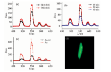
将以下三种情况生长的Si纳米线作为基质进行Tb3+掺杂: (1)以抛光表面为衬底生长的Si纳米线分布较为均匀、 密度较大且生长方向一致, 而以“ 金字塔” 织构表面为衬底生长的Si纳米线分布复杂、 方向杂乱; (2)生长时间分别为15, 30和60 min的Si纳米线(即不同长度); (3)Si纳米线密度为108和1010 cm-2。 室温下, 测试了样品的PL特性及其绿光发射, 结果如图1所示。
 | 图1 Si纳米线对SiNWs:Tb3+ PL特性的影响 (a): 不同分布; (b): 不同长度; (c): 不同密度; (d): 绿光发射Fig.1 Effects of Si nanowires on PL properties of SiNWs:Tb3+ (a): Different distribution; (b): Different length; (c): Different density; (d): Green light emission |
结果显示, 样品实现了较强的绿光发射。 另外, Si纳米线分布范围较大、 密度较小、 长度适中时Tb3+绿光发射强度较大。 因为Si纳米线的密度较大、 较长时, 纳米线交错缠绕, 在一定程度上阻碍稀土粉末和纳米线的充分接触, 使得大量的稀土粉末无法与Si纳米线反应, 导致Si纳米线中的Tb3+浓度较低, 故发光强度弱。
理想的荧光标记物应具有光学稳定性、 良好的光发射特性以及安全无毒不影响生物组织自身功能。 实验测试了样品的温度稳定性、 酸碱稳定性、 抗光漂白能力以及水溶性和分散性。
2.2.1 温度稳定性
荧光材料的温度猝灭性能对材料发光能力和稳定性有着重要的影响。 为了测试样品的光发射强度随着温度变化的关系, 对其进行了不同温度(300~500 K)的PL特性测试, 结果如图2。
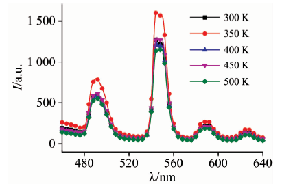 | 图2 样品的温度稳定性Fig.2 Temperature stability of the sample |
结果显示, Tb3+掺杂Si纳米线具有良好的温度稳定特性, 温度升高到500 K时, 发光强度仅降低了8.9%左右。
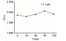
2.2.2 抗光漂白性
光漂白是在光照条件下使发光材料发生化学反应或是结构改变, 而失去荧光发射的特性。 利用功率为450 W、 波长为365 nm的紫外灯对样品分别照射30, 60, 90和120 min, 然后测试其荧光发射特性, 结果如图3所示。
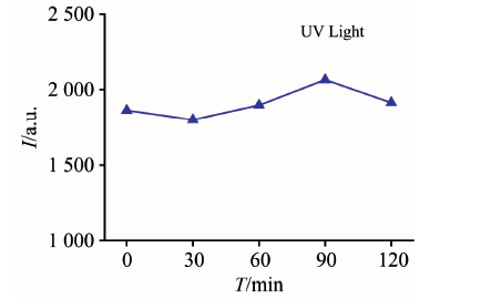 | 图3 样品的抗光漂白特性Fig.3 Anti-photobleaching properties of the sample |
结果显示, 在0~120 min的紫外光照射下, 样品的发光强度几乎没有发生改变。 表明该方法制备的荧光纳米材料具有良好的抗光漂白能力。
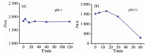
2.2.3 酸碱稳定性
室温下, 测试了样品在强酸和强碱溶液中的稳定性。 首先, 利用HCl溶液配置了pH 1的强酸性溶液, 室温下将样品置于HCl溶液中0~120 min; 然后, 用去离子水冲洗、 烘干, 测试其PL特性, 如图4(a)所示。 可以看出, 在经过120 min的强酸处理后, Tb3+的发光强度没有产生衰减。
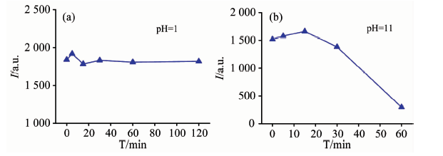 | 图4 样品的酸碱稳定性 (a): 酸; (b): 碱Fig.4 The pH stability of the sample (a): Acid; (b): Alkali |
图4(b)示出了样品在pH 11的NaOH溶液中的稳定性。 样品在15 min之内保持了良好的稳定性; 随后, 发光强度出现了缓慢下降的趋势, 在60 min后, 样品的发光强度变得极其微弱。 分析认为, 在SiNWs:Tb3+表面有一层SiO2包覆层, 而NaOH溶液容易和SiO2发生化学反应, 随着时间延长SiO2层被破坏, 故样品发光强度降低。
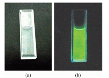
2.2.4 水溶性和分散性
作为荧光标记物要具有良好的水溶性和分散性。 将制备的样品溶于纯净水中振荡120 min; 常温下放置24 h, 将不溶于水的沉淀物过滤掉; 然后, 将溶液置于比色皿中, 分别在波长为365 nm的紫外灯和自然光下观察样品发光特性, 结果分别如图5(a)和(b)所示。
 | 图5 样品的水溶性、 分散性及其发光特性 (a): 自然光; (b): 紫外光Fig.5 Water solubility, dispersity and luminescent of the sample (a): Natural light; (b): Ultraviolet |
可以看出, 溶液无色透明且无沉淀。 紫外光下, 样品发射出了绿色可见光, 且颜色分布比较均匀、 无局部过亮的现象。 表明Tb3+掺杂的Si纳米线具有良好的水溶性和分散性; 另外, 溶液放置30 d后观察未见沉淀物, 因此适合作为荧光标记物。
实验定性研究了不同Si纳米线分布、 长度、 密度对SiNWs:Tb3+绿光发射的影响, 进而测试了样品的光学稳定性。 结果表明, 在衬底为“ 金字塔” 织构表面上、 生长时间为30 min、 以Au为金属催化剂等条件下制备的Si纳米线为Tb3+掺杂衬底时, SiNWs:Tb3+的绿光发射强度较大, 其发光强度峰值位于554 nm(5D4→ 7F5)。 另外, 样品在温度升高到500 K时, 绿光发射强度仅降低了8.9%左右; 用波长为365 nm、 功率为450 W的紫外光源照射120 min, 样品的光发射强度无衰减; 酸、 碱稳定性好, 在pH 1的强酸(HCl)溶液中120 min未见衰减, 在pH 11的强碱(NaOH)溶液中15 min后开始快速衰减; 样品溶于水中放置30天未见沉淀物, 发光亮度均匀且分散性和水溶性较好, 使其作为荧光标记物具有一定的应用价值。
The authors have declared that no competing interests exist.
| [1] |
|
| [2] |
|
| [3] |
|
| [4] |
|
| [5] |
|
| [6] |
|
| [7] |
|
| [8] |
|
| [9] |
|