

小鼠小梁细胞与肌成纤维细胞的同步辐射红外显微光谱
[何明霞1  , 田甜
, 田甜2 , 刘立媛* , 步绍翀, 东莉洁, 张欣欣, 张洪桢]
, 田甜, 步绍翀, 东莉洁, 张欣欣, 张洪桢]
|
|


作者简介: 何明霞, 1965年生, 天津大学测试计量技术及仪器国家重点实验室教授 e-mail: hhmmxx@tju.edu.cn
原发性开角型青光眼是常见的致盲性眼部疾病。 眼压升高是原发性开角型青光眼发生和发展最主要的危险因素, 是由小梁网途径的房水外流排出系统发生病变、 房水流出阻力增加所致。 研究表明, 房水中存在的转化生长因子-β能够使小梁细胞纤维化, 诱导小梁细胞过度增殖, 从而阻碍房水外流, 导致原发性开角型青光眼的发生。 原发性开角型青光眼发病隐蔽, 病程进展缓慢, 早期没有任何症状, 往往到晚期视力视野有显著损害时, 才会被发现, 因此原发性开角型青光眼的早期诊断尤为重要。 同步辐射红外显微成像结合高亮度、 高分辨率的同步辐射源, 同时配备傅里叶变换红外光谱仪与红外显微镜, 可以实现细胞的检测。 这对从分子层面获取细胞的变化信息, 深入理解疾病的发病机制以及疾病的早期诊断具有非常重要的意义。 虽然有很多红外光谱在生物医学领域的研究报道, 但是应用红外光谱显微成像技术研究细胞等生物医学体系仍然是亟待发展的领域, 并且目前未找到关于红外光谱用于小梁网细胞的检测报道。 在体外用转化生长因子-β对老鼠小梁网细胞进行诱导, 使其转化为肌成纤维细胞, 模拟小梁细胞纤维化过程。 对小梁网细胞以及经转化生长因子-β诱导形成的肌成纤维细胞进行同步辐射红外显微成像及光谱分析, 并进一步探讨同步辐射用于早期诊断原发性开角型青光眼的可行性。 研究表明肌成纤维细胞内的弹性蛋白明显高于小梁网细胞, 而弹性蛋白中95%为非极性氨基酸, 即氨基酸的侧链基团R基只有C和H两种元素。 对比两种细胞的红外谱图, 发现在2 934, 2 900和2 845 cm-1, 肌成纤维细胞的 CH3, CH2和CH的伸缩振动明显强于小梁网细胞, 推测可能是由于转化生长因子-β诱导后细胞内弹性蛋白增加所致。 在细胞层面检测了小梁网细胞的过度增殖, 为将来可以直接获取细胞的红外光谱从而检测小梁网细胞的增殖程度, 进而检测原发性开角型青光眼等疾病奠定了基础。 得出同步辐射红外谱学与显微成像有望成为检测原发性开角型青光眼新手段的结论, 也为将来便携式红外显微光谱仪临床实时检测青光眼等疾病提供了依据。
Primary open angle glaucoma is a common blind eye disease. Elevated intraocular pressure is the most important risk factor for the occurrence and development of primary open-angle glaucoma. It is caused by the lesion of aqueous humor outflow system and the increase of aqueous humor outflow resistance in trabecular meshwork pathway. It has been shown that TGF- β in aqueous humor can make trabecular cells fibrosis and induce excessive proliferation of trabecular cells, thus hindering the outflow of aqueous humor, leading to the occurrence of primary open-angle glaucoma. POAG is a covert disease with slow progression and no symptoms in the early stage. It is often found only when the visual field is significantly impaired in the late stage. Therefore, the early diagnosis of it is particularly important. Synchrotron radiation infrared microimaging combined with high luminance and high resolution synchrotron radiation source with Fourier transform infrared spectrometer and infrared microscope can realize cell detection. It is very important to obtain cellular change information from molecular level and to understand the pathogenesis of disease and the early diagnosis of disease. Although there are many research reports on infrared spectroscopy in biomedical field, the application of infrared spectroscopy microscopic imaging technology to the study of biomedical systems such as cells is still an area in urgent need of development. At present, no infrared spectroscopy has been found for the detection of trabecular meshwork cells. In this paper, TGF-β was used to induce rat trabecular meshwork cells into myofibroblasts in vitro, simulating the process of trabecular meshwork cells fibrosis. The meshwork cells and the myofibroblasts induced by TGF-β were studied by synchrotron radiation infrared microscopy and spectral analysis, and the feasibility of using synchrotron radiation in the early diagnosis of primary open-angle glaucoma was discussed. Study have shown that the elastin in myofibroblasts was significantly higher than that in trabecular meshwork cells, and 95% of elastins were nonpolar amino acids. Comparing the IR spectra of the two kinds of cells, it was found that the stretching vibration of CH3, CH2 and CH of myofibroblasts at 2 934, 2 900 and 2 845 cm-1 was stronger than that of trabecular meshwork cells, which might be due to the increase of intracellular elastin induced by TGF-β. In this paper, we detected the excessive proliferation of trabecular meshwork cells at the cellular level, which laid a foundation for obtaining the infrared spectrum of cells directly and detecting the proliferation of trabecular meshwork cells in the future, and then for detecting diseases such as primary open angle glaucoma. It is concluded that synchrotron radiation infrared spectroscopy and microscopic imaging are expected to be new methods for detecting POAG and provide a basis for real-time clinical detection of glaucoma by portable infrared microspectrometer.
原发性开角型青光眼(primary open angle glaucoma, POAG)是常见的致盲性眼部疾病。 眼压升高是原发性开角型青光眼发生和发展最主要的危险因素, 是由小梁网途径的房水外流排出系统发生病变、 房水流出阻力增加所致[1, 2]。 研究表明, 房水中存在的转化生长因子-β (transforming growth factor-β , TGF-β )能够使小梁细胞纤维化[3, 4], 诱导小梁细胞过度增殖, 从而阻碍房水外流, 导致原发性开角型青光眼的发生。 原发性开角型青光眼发病隐蔽, 病程进展缓慢, 早期没有任何症状, 往往到晚期视力视野有显著损害时, 才会被发现, 因此原发性开角型青光眼的早期诊断尤为重要。 同步辐射红外显微成像结合高亮度、 高分辨率的同步辐射源, 同时配备傅里叶变换红外光谱仪与红外显微镜, 可以实现细胞检测。 虽然生物医学领域有很多关于红外光谱的研究报道, 但是红外光谱显微成像技术在细胞等生物医学领域的研究应用仍然是极需迫切发展, 目前并未找到关于红外光谱用于小梁网细胞的检测报道。 因此用同步辐射红外显微成像研究与原发性开角型青光眼等疾病有关的小梁网细胞, 从分子层面获取细胞的变化信息, 了解疾病的发病机制和疾病的早期诊断非常重要。
本工作的总体目标是通过研究小梁网细胞及肌成纤维细胞的红外光谱及成像, 探究同步辐射红外显微成像与红外光谱诊断原发性开角型青光眼的可行性, 并探索两种细胞红外光谱差异的机制。 具体目标是: (1)获得小梁网细胞以及经TGF-β 诱导后的肌成纤维细胞的红外光谱与成像; (2)研究两种细胞红外谱图的差异, 探索产生差异的机制; (3)讨论红外谱学与显微成像用于诊断小梁网细胞的过度增殖, 进而诊断原发性开角型青光眼的可能性。
取材前, 用紫外线对实验室、 超净工作台消毒30分钟。 在无菌条件下将小鼠小梁网细胞(rat trabecular meshwork cells, RTMC)以1× 10⁶密度爬片于6孔细胞培养板中预先放置的盖玻片上, 待细胞汇合度约70%, 依据实验分组进行相应的药物刺激处理; 正常细胞组: 常规培养的RTMC细胞; TGF-β 组: 用10 ng· mL-1的TGF-β 刺激细胞; 24 h后, 终止2组细胞的培养, 滴入4%多聚甲醛室温10 min进行固定。
上海光源(Shanghai synchrotron radiation facility, SSRF)[5]是我国建造的第三代中能同步辐射光源, 其能量目前居世界第四, 性能超过同能区现有的其他第三代同步辐射光源。 上海光源的时间分辨与谱学显微红外光束线站的光源亮度优于传统光源亮度2~3个数量级[6, 7], 具有高通量、 高信噪比等优势。 测试分辨率可达5 μ m。 而一般细胞大小为10~20 μ m, 因此可以深入研究细胞。 高度集成的操作环境, 大大提升了实验效能。
本实验样品分为两组, 小梁网细胞组为对照组, 肌成纤维细胞组为实验组。 每组选取10个细胞生长状况良好的区域进行微区成像, 将每个区域中心点位置的红外光谱作为两种细胞的红外谱图, 观察10次获得的小梁网细胞的红外光谱, 具有很好的一致性。
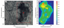
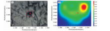
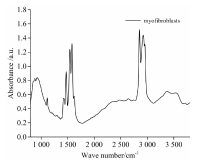
如图1(a)为小梁网细胞在红外显微镜下的光学图像, 图中红色框内区域为扫描区域, 扫描面积为20 μ m× 25 μ m; 图1(b)为红外显微光谱成像图, 图中不同颜色代表不同的吸光度(吸光度值见图1(b)右侧条状区域), 蓝色部分为空白氟化钡窗片区域, 绿色部分为样品区域; 图2为小梁网细胞的红外光谱图。 图3(a)为肌成纤维细胞在红外显微镜下的光学图像, 图中红色框内区域为扫描区域, 扫描面积为20 μ m× 25 μ m; 图3(b)为红外显微光谱成像图, 图中不同颜色代表不同的吸光度(吸光度值见图3(b)右侧条状区域); 图4为肌成纤维细胞的红外谱图。
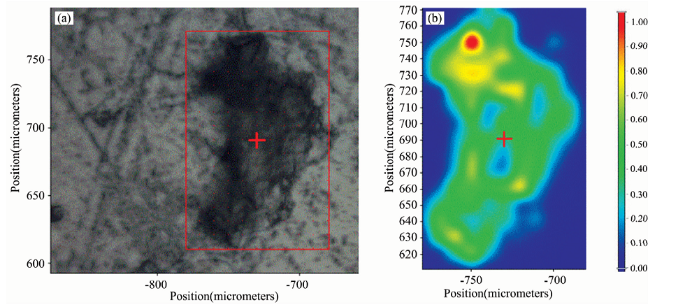 | 图1 (a) 红外显微镜下小梁网细胞光学图; (b) 小梁网细胞红外显微成像图Fig.1 (a) Trabecular meshwork cells under infrared microscope; (b) Infrared spectral imaging of trabecular meshwork cells |
 | 图2 小梁网细胞红外光谱图Fig.2 Infrared spectrum of trabecular meshwork cells |
 | 图3 (a) 红外显微镜下肌成纤维细胞光学图; (b) 肌成纤维细胞红外显微成像图Fig.3 (a) Image of myofibroblasts under infrared microscope; (b) Infrared spectral imaging of myofibroblasts |
 | 图4 肌成纤维细胞红外光谱图Fig.4 Infrared spectra of myofibroblast |
小梁网细胞与诱导后的肌成纤维细胞的红外光谱的主要峰及其位置归属如表1所示。
| 表1 小梁网细胞与肌成纤维细胞的红外光谱的主要峰及其位置归属 Table 1 Main peaks and location of infrared spectra of trabecular meshwork cells and myofibroblasts |
由表1可知, 小梁网细胞经TGF-β 诱导后, 糖类O— H的伸缩振动、 糖类C— O— C的伸缩振动、 烷烃类化合物基团CH3, CH2和CH的伸缩振动明显增强。 即细胞中糖类、 烷烃类化合物增多。 研究表明[11, 12], 小梁网细胞经转化生长因子-β 诱导后, 细胞中弹性蛋白会显著增加。 弹性蛋白的增多会加重细胞外基质堆积, 房水引流阻力增加, 从而导致原发性开角型青光眼的发生。 而弹性蛋白中95%为非极性氨基酸, 与实验结果中CH3, CH2和CH的伸缩振动增强相符。
小梁网细胞经TGF-β 诱导为肌成纤维细胞后, 细胞中糖类、 蛋白、 脂类等的结构与含量发生了变化, 各种化学组成及其振动方式也发生了改变, 导致诱导前后细胞的红外显微光谱图有着明显差异。 由于肌成纤维细胞的弹性蛋白增多, 使得在2 934, 2 900和2 845 cm-1的CH3, CH2和CH的伸缩振动明显强于小梁网细胞。 因此可以通过监测2 934, 2 900和2 845 cm-1 下的峰强来区分小梁网与肌成纤维细胞, 进而检测原发性开角型青光眼等疾病。
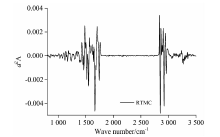
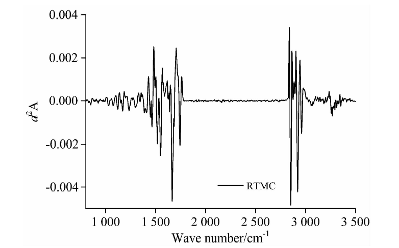
目前导数光谱已经在红外光谱分析中得到了广泛应用[13]。 如果初始红外谱的吸光度很小, 信号质量会严重损失。 若样品的吸收峰重叠, 也会干扰光谱信号的分析。 二阶导数光谱不仅能消除基线的影响, 同时可以降低背景干扰, 得到更完备的样品信息。 用二阶导数光谱法处理小梁网细胞和肌成纤维细胞的红外光谱数据, 并用Savitzky-Golay平滑方法处理, 得到两种细胞的二阶红外谱。 分别使用了7点、 9点、 11点、 13点的平滑模型, 结果显示9点的平滑结果较好, 如图5为波数范围在3 800~1 000 cm-1范围内小梁网细胞的红外二阶导数光谱, 图6为波数范围在3 800~1 000 cm-1范围内肌成纤维细胞的红外二阶导数光谱。
 | 图5 小梁细胞的二阶导数光谱Fig.5 Second derivative spectra of trabecular cells |
 | 图6 肌成纤维细胞的二阶导数光谱Fig.6 Second derivative spectra of myofibroblast |
如图5, 在肌成纤维细胞的二阶导数光谱的下凸谷位置3 289, 3 066, 2 963, 2 922, 2 853, 1 742, 1 667, 1 546, 1 459, 1 401, 1 300, 1 246, 1 170, 1 121以及1 0811 cm-1分别对应原光谱吸收峰的位置, 与原光谱基本吻合。 如图6, 在肌成纤维细胞的二阶导数光谱的下凸谷位置2 957, 2 934, 2 900, 2 845, 1 620, 1 587, 1 539, 1 464, 1 434以及1 135 cm-1分别对应原光谱吸收峰的位置, 与原光谱基本吻合。
细胞是最基本的生命系统, 是生物体结构和功能的基本单位。 深入的研究细胞是阐释生命、 探究疾病机理的关键。 组织层面的疾病检测, 往往具有延迟性, 当组织层面发生病变时, 很多生物细胞可能早已发生坏死、 癌病等情况。 因此对细胞的深入研究对于疾病的早期诊断和发病机制的探索有着深远意义。 以往受限于仪器的发展, 红外技术用于细胞的研究较少, 但是随着仪器的发展, 测量分辨率不断提高, 将组织层面的疾病研究扩展到细胞层面成为可能。 本文从小梁细胞出发, 研究小梁细胞和病理状态下的肌成纤维细胞的红外光谱, 对于诊断原发性开角型青光眼等眼部疾病有着重要的作用, 同时对于眼部疾病乃至其他疾病致病机理的探索提供了依据和方向。
原发性开角型青光眼是生活中常见的眼部疾病。 眼压升高是原发性开角型青光眼发生和发展最主要的危险因素, 是由小梁网途径的房水外流排除系统发生病变、 房水流出阻力增加所致。 TGF-β 是多肽形式的一组细胞因子, 能够使小梁网细胞纤维化, 导致小梁网过度增殖。 而小梁细胞的过度增殖就会导致原发性开角型青光眼。 本章用TGF-β 诱导体外培养的小鼠小梁网细胞, 模拟小梁细胞纤维化过程。 小梁网细胞为对照组, 诱导后的肌成纤维细胞为实验组。 结合高亮度、 高分辨率的同步辐射光, 对两组细胞进行红外显微成像。 从化学键和官能团的振动模式分析了小梁细胞和肌成纤维细胞的红外光谱。 用Savitzky-Golay方法处理两种细胞的二阶导数光谱, 结果显示二阶导数谱与原光谱吻合很好。 实验发现在2 934, 2 900和2 845 cm-1 下, 肌成纤维细胞的CH3, CH2和CH的伸缩振动明显强于小梁网细胞, 猜测可能是由于小梁网细胞经TGF-β 诱导后弹性蛋白含量增多, 而弹性蛋白中95%为非极性氨基酸使得CH3, CH2和CH的伸缩振动增强。 因此同步辐射红外显微成像有望成为检测原发性开角型青光眼的新手段, 也为将来便携式红外显微光谱仪临床实时检测青光眼等疾病提供了依据。
致谢: 感谢天津医科大学眼科医院对样品的支持, 以及上海蛋白质科学研究所和上海同步辐射装置红外谱学与显微成像(BL01B)线站全体工作人员。
| [1] |
|
| [2] |
|
| [3] |
|
| [4] |
|
| [5] |
|
| [6] |
|
| [7] |
|
| [8] |
|
| [9] |
|
| [10] |
|
| [11] |
|
| [12] |
|
| [13] |
|