
Chemical Analysis of Hydroxyapatite Artificial Bone Powders by Energy Dispersive X-Ray Fluorescence Spectrometry (EDXRF)
引用本文
O. K. Koksal, G. Apaydin, E. Cengiz, L. Samek, I˙.H. Karahan, A. Tozar, M. Lankosz. Chemical Analysis of Hydroxyapatite Artificial Bone Powders by Energy Dispersive X-Ray Fluorescence Spectrometry (EDXRF). Spectroscopy and Spectral Analysis, 2018,38(8): 2645-2649. 
Doi: 10.3964/j.issn.1000-0593(2018)08-2645-05
Permissions
Copyright©2018, 光谱学与光谱分析
光谱学与光谱分析 所有
Chemical Analysis of Hydroxyapatite Artificial Bone Powders by Energy Dispersive X-Ray Fluorescence Spectrometry (EDXRF)
Abstract
Stoichiometric hydroxyapatite (HA) nanoparticles were synthesized by a wet chemical method. Calcium nitrate tetra hydrate used as calcium source and dibasic ammonium phosphate used as phosphorous source. Calcium nitrate tetra hydrate and dibasic ammonium phosphate solutions were prepared by dissolving the salts in distilled water. Stoichiometric hydroxyapatite nanoparticles used by artificial bone powders and synthesized by a wet chemical method were analyzed using EDXRF method.The concentrations of K, Ca, Ti, V, Cr, Fe, Ni, Cu, Sr and Pb for artificial bone powders have been determined. Besides, Calcium contents were evaluated according to the agitation time and temperature in the production process.
Key words:
EDXRF; X-ray Tube; Elemental analysis; Hydroxyapatite
Introduction
Most tissues of an organism have hierarchical structure. Also bone and tooth enamel do. This hierarchical structure determines the mechanical properties of bone substantially. Hydroxyapatite (HA) constitutes the bottom level of this hierarchy with its Nano metric size. (HA) (Ca10(PO4)6(OH)2) is an inorganic component which existence is natural in human bone and tooth enamel[1]. HA is primarily composed of calcium and phosphorus with a stoichiometric Ca/P ratio of 1.667[2]. Pure HA mainly crystallizes in the monoclinic space group P21/b. Nevertheless, a phase transition of monolithic to hexagonal (space group P63/m)[3, 4] occurs at the temperatures above 250 ℃[5, 6].
HA has been used as bone cement[7], scaffolds[8] and coating on metal[9] because of its excellent biocompability[10]. Moreover, due to bone bonding properties of HA, it becomes extensively used metal coatings in biomedical applications[11, 13]. As metallic substrate provides the intended mechanical properties, they utilize HA to obtain an osteoconductive surface for new bone growth, immobilizing the metal implant and transferring load to the skeleton, helping to overcome bone atrophy problem[14, 17]. Even for no-load-bearing applications (such as tooth root substitutes, cystic cavities, filling of periodontal pockets and spinal fusions etc.) adequate mechanical properties, such as hardness, bend strength, fracture toughness, and wear resistance, are still essentials.
It is well known that mechanical properties of ceramics are anchored in grain size[18, 19]. As diameter of ceramic particle decreases, the strength of the ceramic increases[20, 21, 22]. Grain size is a key factor not only for mechanical properties of ceramics but also for many physical properties. Furthermore, decreased grain size means increased surface area. In some applications (drug delivery systems or corrosion coatings etc.) increased surface area and controlled morphology are quite important.
Pervasive methods including wet chemical precipitation[23, 24], hydrothermal[25, 26], hydrolysis[27, 28], mechanochemical[29, 30], sol-gel[31, 32], and electrochemical[33, 34] have been used to fabricate Nano-hydroxyapatite powder. Wet chemical precipitation method looks promising due to its ease of perform, low cost and homogeneity in particle size distribution. Recent studies showed that the reactivity of chemical species in solution, involved in a synthesis process can be stimulated by ultrasonic agitation the wet chemical precipitation solution. This kind of agitation can accelerate heterogeneous reactions between liquid and solid reactants efficaciously by a phenomena called cavitation which can be characterize as simultaneous formation, growth and collapse of micro-bubbles occurring[35]. Cavitation can be stimulated by this kind of intense irradiation process and causes to dissolution-precipitation of solids which reduces particle size and surface activation of the product[36, 38].
In the present work, Nano-sized HA powders were synthesized via ultrasonically assisted wet chemical precipitation method using a very simple solution containing calcium nitrate and ammonium phosphate. Effect of ultra-sonication time was investigated and compared with magnetic stirring conditions. Additionallychemical concentrations of Nano-sized HA powders synthesized via ultrasonically assisted wet chemical precipitation method using a very simple solution containing calcium nitrate and ammonium phosphate were determined using EDXRF technique. Besides, it was researched how the calcium content changes with the agitation time and temperature.
1 Material and methods
1.1 Sampling
Stoichiometric HA nanoparticles were synthesized by a wet chemical method. Calcium nitrate tetra hydrate (Ca(NO3)2· 4H2O) used as calcium source and dibasic ammonium phosphate ((NH4)2HPO4) used as phosphorous source. Equal volumes of 1.0 mol· L-1 calcium nitrate tetra hydrate and 0.6 mol· L-1 dibasic ammonium phosphate solutions were prepared by dissolving the salts in distilled water. Ammonium phosphate solution was very slowly added to calcium nitrate solution dropwise at a rate of 5 mL· min-1. Rapid sedimentation of a white powder was seen while adding the solutions to each other. To avoid this issue calcium nitrate solution magnetically stirred through the wet chemical precipitation. pH of the final solution was regulated to 10 with ammonia NH3· H2O and then to 11 with NaOH. All these procedures were carried out at room temperature. The final solution was subjected to different stirring conditions shown in Table 1.
| Table 1 The conditions of sample preparation |
And all of them aged at room temperature for 24 h. After aging, the solutions were washed with deionized water to acquire neutrality and then washed with mixture of ethanol-methanol. Finally, the samples filtered with filter paper and then dried in a drying oven and then dried samples grinded in a muller.
1.2 Chemical Analysis
Concentrations of the following elements were quantified: K, Ca, Ti, V, Cr, Fe, Ni, Cu, Sr and Pb. Samples of artificial bone were analyzed with the use of a multifunctional energy dispersive X-ray fluorescence spectrometer as medium samples. They were mixed with cellulose and then prepared as pellets. The instrument is a micro-beam X-ray fluorescence spectrometer with capillary X-ray optics, a broad X-ray beam from a molybdenum secondary target for XRF analysis of bulk samples and a total reflection X-ray technique. The molybdenum tube is the source of X-rays. The tube has the power of 2 kW. The excited X-rays were detected by a Si(Li) detector with resolution of 170 eV at an energy of 5.9 keV. Data collection was completed using the Canberra system. The measurements were carried out under the following conditions: voltage of 55kV, current of 30 mA, measurement time of 1 000 s, under atmospheric air. In order to calculate the concentrations of different elements in the bones, the spectrometer was calibrated using standard pellets with known concentration of elements. The calibration was verified by the analysis of the NIST Standard Reference Material Soil-7. The XRF spectra were quantitatively analyzed with the use of the QXAS package[39].
2 Results and Discussions
In this study, HA were produced by wet chemical method as well as elemental concentrations of artificial bone powders were determined. Following elements were measured by EDXRF method: K, Ca, Ti, V, Cr, Fe, Ni, Cu, Sr and Pb.Here, Ti, V, Cr, Fe, Ni, Cu, Sr and Pb elements are impurities in artificial bone powders thought to arise from the manufacturing process. Results of elemental analysis forthese powders are illustrated in Figure 1 and Table 2, respectively.
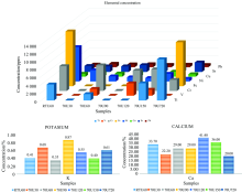
 | Fig.1 Elemental concentrations of artificial bone powders |
| Table 2 Results of elemental EDXRF analysis of artificial bone powders |
The calcium concentration is the highest concentration values for all samples as percentage. It is followed by a lower value potassium as a percent. Strontium, lead, titanium, vanadium, chromium, iron, copper, and nickel have elemental concentrations as ppm (part per million). Strontium and lead concentrations have values close to each other in entire specimens. Titanium, vanadium, chromium and iron could not be detected in some samples while determining in some samplings.
70U720 has the lowest concentrations of Ca, Ni, Cu, Pb in all of the samples but it has the highest concentration of Sr in entire samples and not detected vanadium. 70U90 has the highest concentration of K in all of the samples. 70U30 has the highest concentration of Pb in all of the samples. Titanium, vanadium, chromium, and iron have not been determined in 70U90 and 70U30. RTU60 and 70U720 has the highest concentrations of Fe in the samples but Fe was not dedicated in the rest of the samples. 70U60 has the lowest concentration of K among all samples.
Calcium was found between 20%~42% in entire samples. Potassium is between 0.35%~0.87% in all of the specimens. Titanium was determined among 16~103 ppm but could not been determined in 70U120, 70U30, 70U90. Vanadium was found between 15~31 ppm but could not been detected in RTU60, 70U30, 70U90 and, 70U720. Chromium was detected between 27~62 ppm but was not found in 70U30 and 70U90. Iron has 110 and 137 ppm concentration values but it could not been found in rest of the samples. Nickel was between 8~29 ppm in the specimens. Concentrations of copper had values from 8 ppm to 13 ppm. Strontium existed between 14~24 ppm in the samples and the concentration levels are almost same value except the highest value in the sample. Concentration values of lead observed in the samples are almost close to each other. Concentrations of lead are between 6~9 ppm.
One of the main purposes of this paper is to determine Ca concentration and is to observe how it changeswith production parameters. Ca/P ratio was adjusted to be 1.67 in all samples. The hydroxyapatite (HA) which the Ca/P is 1.67 is the most important calcium orthophosphate in vertebrates such as humans. The mineral structure of human bone mainly consist of Nano-sized HA molecules. Ca/P ratio determines the physical (e.g. tensile strength[40]), biological (e.g. Osteoconductivity[41]), or chemical (e.g. Solubility[42]), properties of bone or other bio ceramic material which is intended to implantation. On account of this, the compositions of calcium and potassium elements play a key role in the production of artificial bone powder. However, the phosphate could not be determined. In the performed measurement, it was observed that calcium contents varied with agitation time and temperature. When Table 1 and Table 2 are examined together, the calcium content increased from 22.2% to 41.4% with rising agitation time from 30 to 120 min at the same temperature. After this time it was found that the calcium content decreased. If agitation change from ultrasonic to magnetic concentration of calcium decreased to 20%. When the agitation time is constant, the calcium content is inversely proportional by temperature and was equal to 33.7% for RTU60 sample and 29% for 70U60 one.For potassium relationship between content and agitation temperature was observed. Higher temperature of agitation caused lowering of content of potassium from 0.41% to 0.35%. The highest concentration of potassium was observed for agitation time equal to 90 min and the lowest concentration of potassium was for agitation timeequal to 60 min at the same temperature.
Wet chemical method can be used for synthesis stoichiometric hydroxyl apatite nanoparticles. Energy dispersive X-ray fluorescence analytical tool seems to be a good elemental analysis technique for analyzing the artificial bone powders since of the advantage of being non-destructive.
The authors have declared that no competing interests exist.
参考文献
| [1] |
|
| [2] |
|
| [3] |
|
| [4] |
|
| [5] |
|
| [6] |
|
| [7] |
|
| [8] |
|
| [9] |
|
| [10] |
|
| [11] |
|
| [12] |
|
| [13] |
|
| [14] |
|
| [15] |
|
| [16] |
|
| [17] |
|
| [18] |
|
| [19] |
|
| [20] |
|
| [21] |
|
| [22] |
|
| [23] |
|
| [24] |
|
| [25] |
|
| [26] |
|
| [27] |
|
| [28] |
|
| [29] |
|
| [30] |
|
| [31] |
|
| [32] |
|
| [33] |
|
| [34] |
|
| [35] |
|
| [36] |
|
| [37] |
|
| [38] |
|
| [39] |
|
| [40] |
|
| [41] |
|
| [42] |
|